
Bringing science to life through photography!

European Researchers' Night 2025 Photo Contest
Welcome to the European Researchers' Night 2025 Photo Contest, a celebration of science through the lens of creativity!
The European Researchers' Night (ERN) is a flagship initiative of the European Commission, running since 2005 under the Marie Skłodowska-Curie Actions. Held simultaneously in over 30 countries and 300 cities across Europe, this event bridges the gap between cutting-edge research and the public, fostering a deeper understanding of how science shapes our world.

It’s time to showcase the creative side of science
Open to everyone
This Year's Theme: Science for a Sustainable Future
Join us in exploring how research addresses the world's most pressing challenges—from environmental protection to economic growth and social equity. This year’s contest invites you to capture the transformative impact of science in everyday life.
Photo credits: Where is Earth expanding to, by Ana Pinheiro

2025 Edition
Tackling global challenges
Explore how research tackles global challenges like environmental protection, economic growth, and social equity. This year’s contest invites you to showcase science's transformative impact on daily life.
Check all the participations and winners below ⤵
2023
A vision of unique perspectives
The #ERN2023 Photo Competition invited everyone to submit up to two original photos under the theme ‘Science for Everyone – Sustainability and Inclusion’.
2022
Creativity was celebrated
#ERN2022 Photo Competition celebrated the fusion of science, technology, and art, sparking diverse conversations and inspiring ideas
Light, lens, and the unknown
Photo Competition Gallery


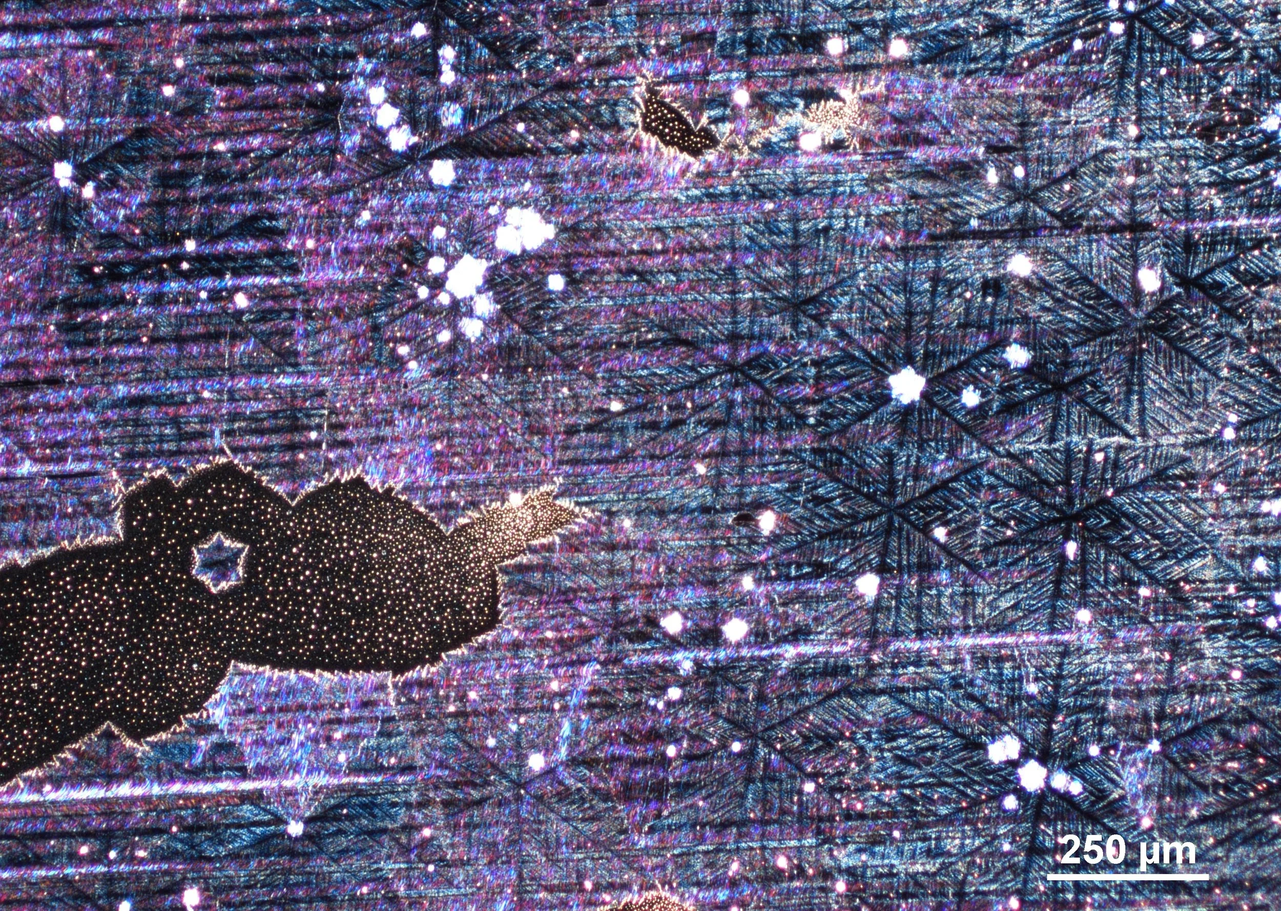
“Microcosmic Chemical Gardens”
This image shows a nucleation region within a crystalline film formed from a droplet on glass. Under crossed polarizers and a wave plate, a floral-like birefringent pattern emerges as interlocking crystal grains solidify. Magnification: 100x.
Captured at The Art of Science Photography studio.
Karl Gaff, with a background in physics and expertise in advanced imaging techniques, captures the hidden beauty of crystals using polarised light microscopy, blending science and art.

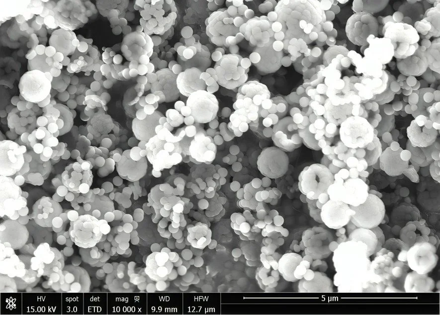
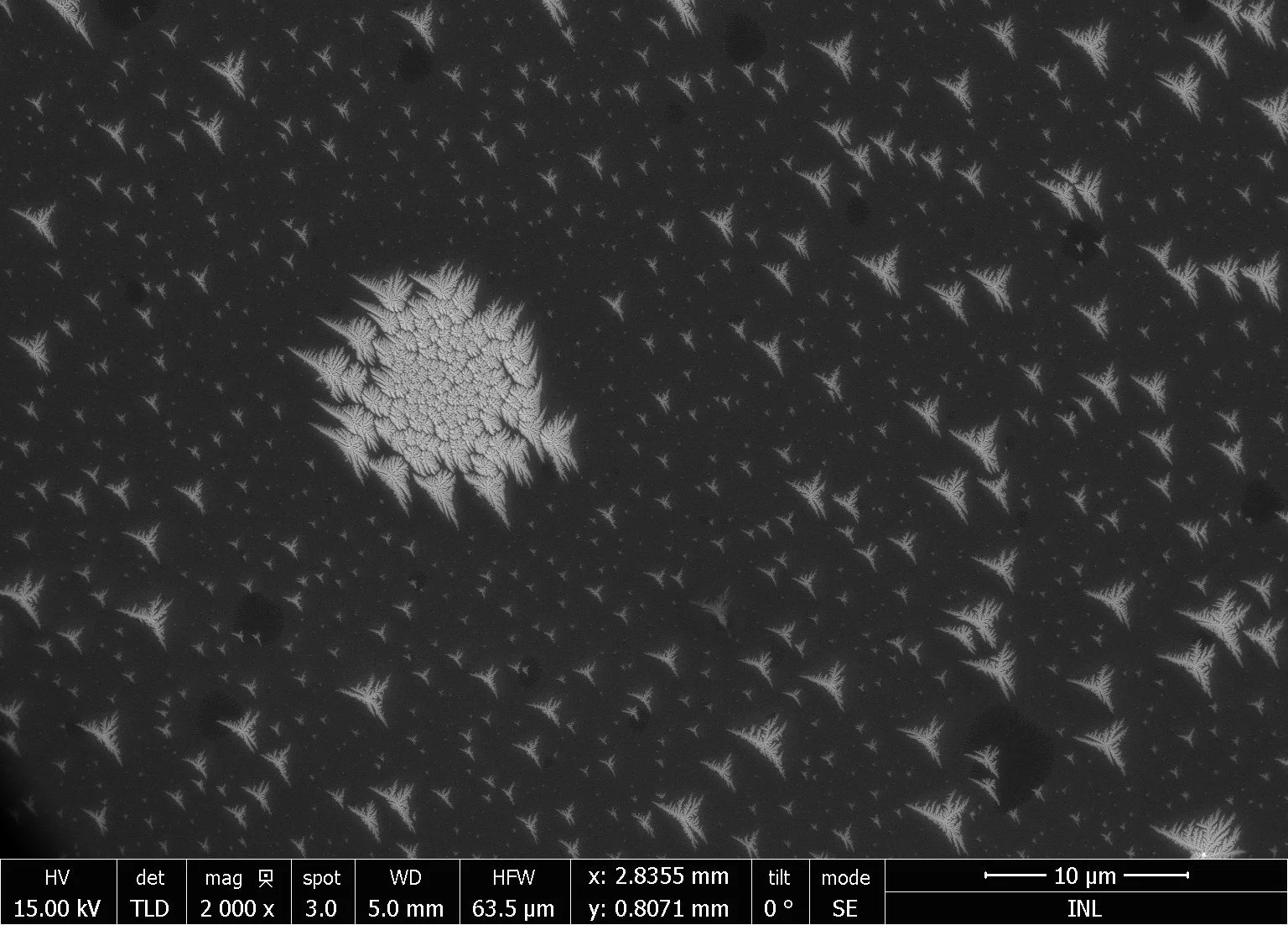
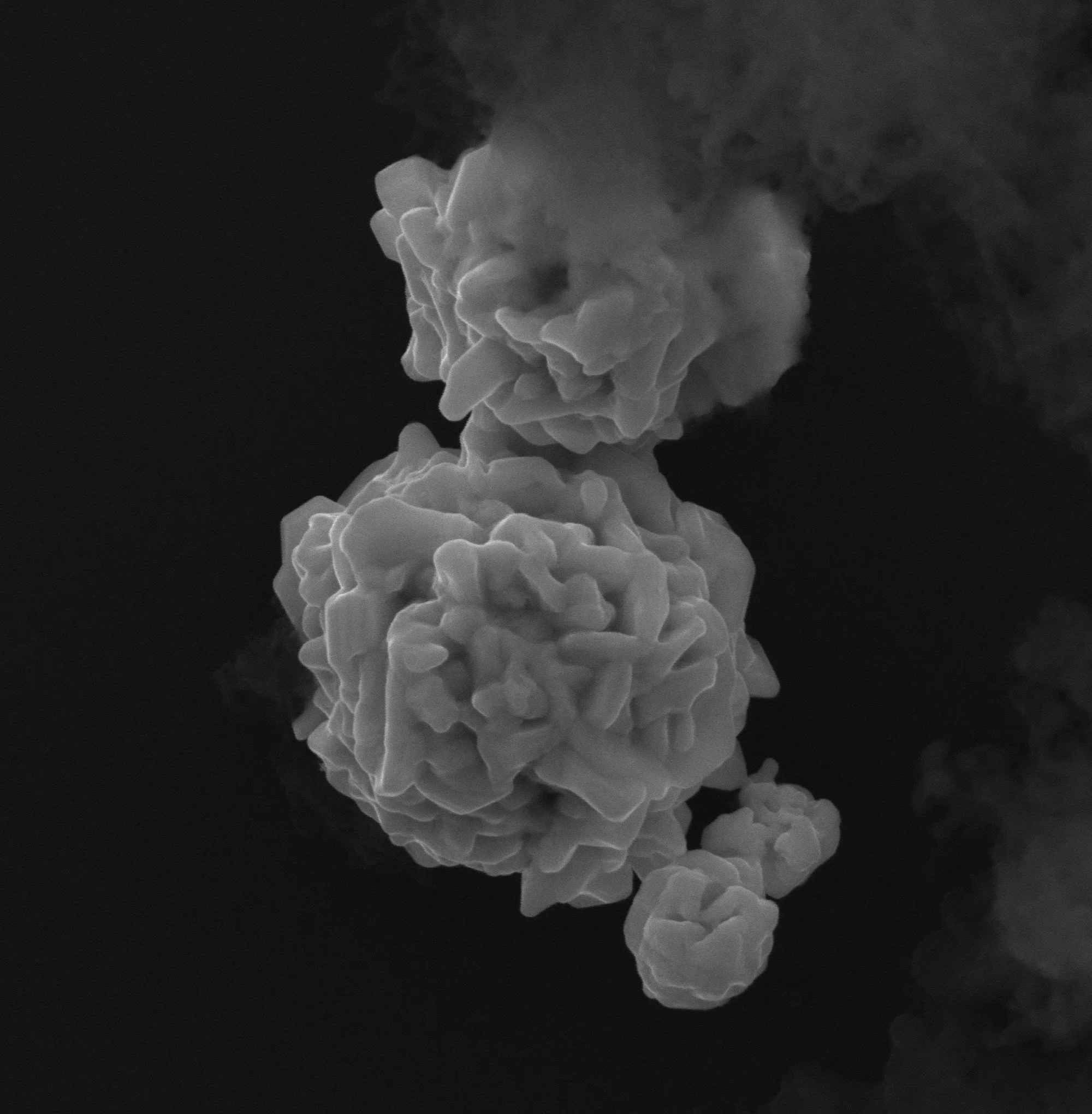
“Hidden Artifacts of Science”
This SEM image shows the intricate surface of a PEG hydrogel, a key material in regenerative medicine and drug delivery. The vivid crystal-like structures are artefacts caused by prolonged electron beam exposure. Captured at Izmir Biomedicine and Genome Center with a Zeiss GeminiSEM 500, the image highlights how research into biodegradable hydrogels is shaping innovation across healthcare and industry, proving science is more than discovery; it’s a dialogue with nature.
Emine Kahraman, Postdoctoral Researcher at MIA Portugal in the Systems Cell Biology Group.

“A New Wave of Science”
SEM image of 4-leaf clover-shaped micropillars made of stacked semiconductors (GaAs, AlGaAs, AlAs), designed as ultra-low power micro-LEDs for neuromorphic computing. Manually colorised: the burgundy silicon nitride layer protects the sample, while the blue gold contact—partially covering the pillars—resembles a flowing wave.
João Azevedo, Research Fellow in the Nieder Research Group at INL, under the supervision of Dr Bruno Romeira, and PhD student at the University of Minho (Braga).

“Sustaining from Above, Earth's Watchful Eyes”
Satellites streak across the night sky, silently shaping our world — from tracking climate change to connecting communities. Captured during the passage of comet C/2023 A3 (Tsuchinshan–ATLAS) over a beach in Sintra, this image reflects the delicate balance between technology and our planet’s future.
João Martins, Molecular Biologist and Photographer passionate about scientific discovery and visual storytelling.
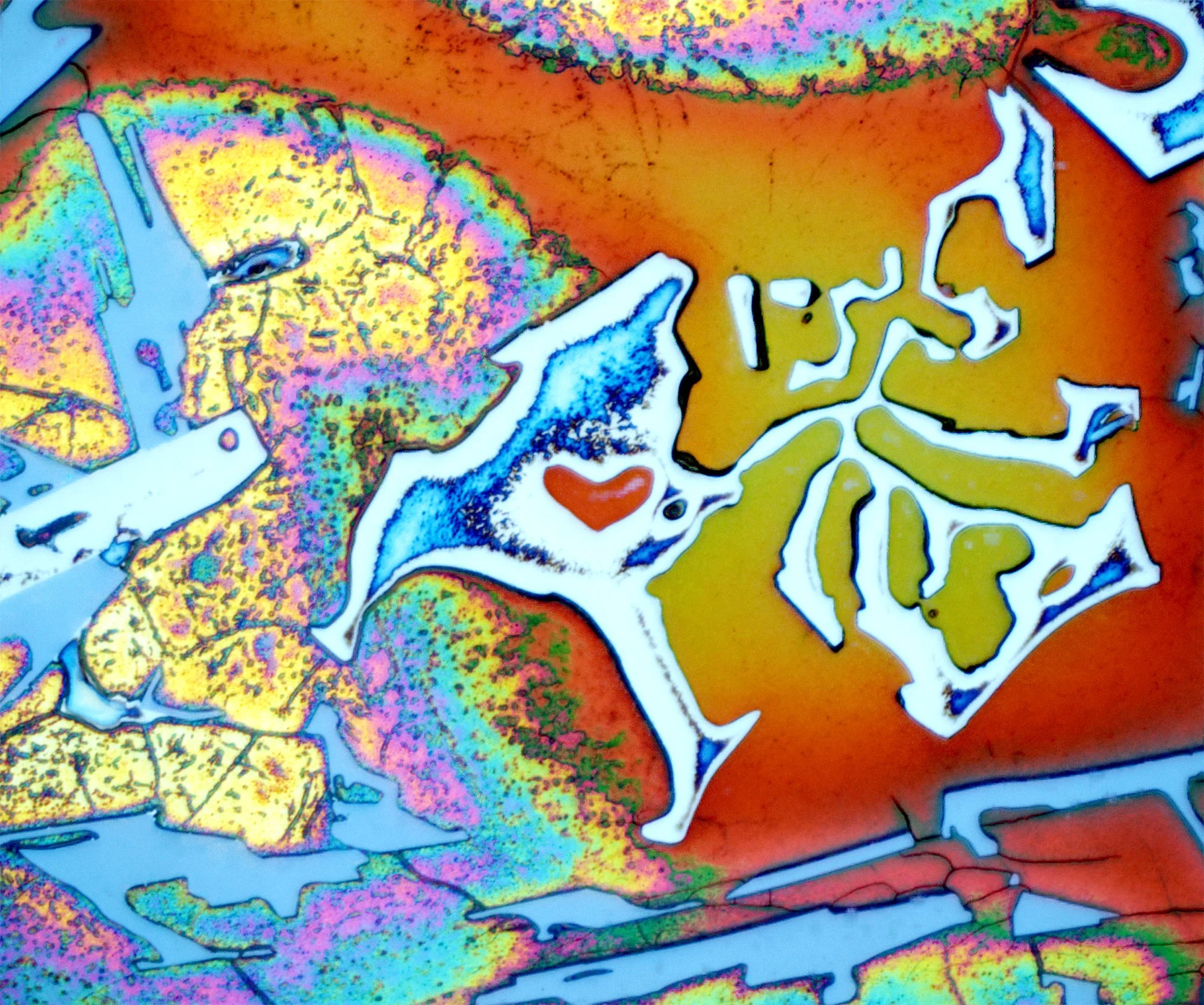
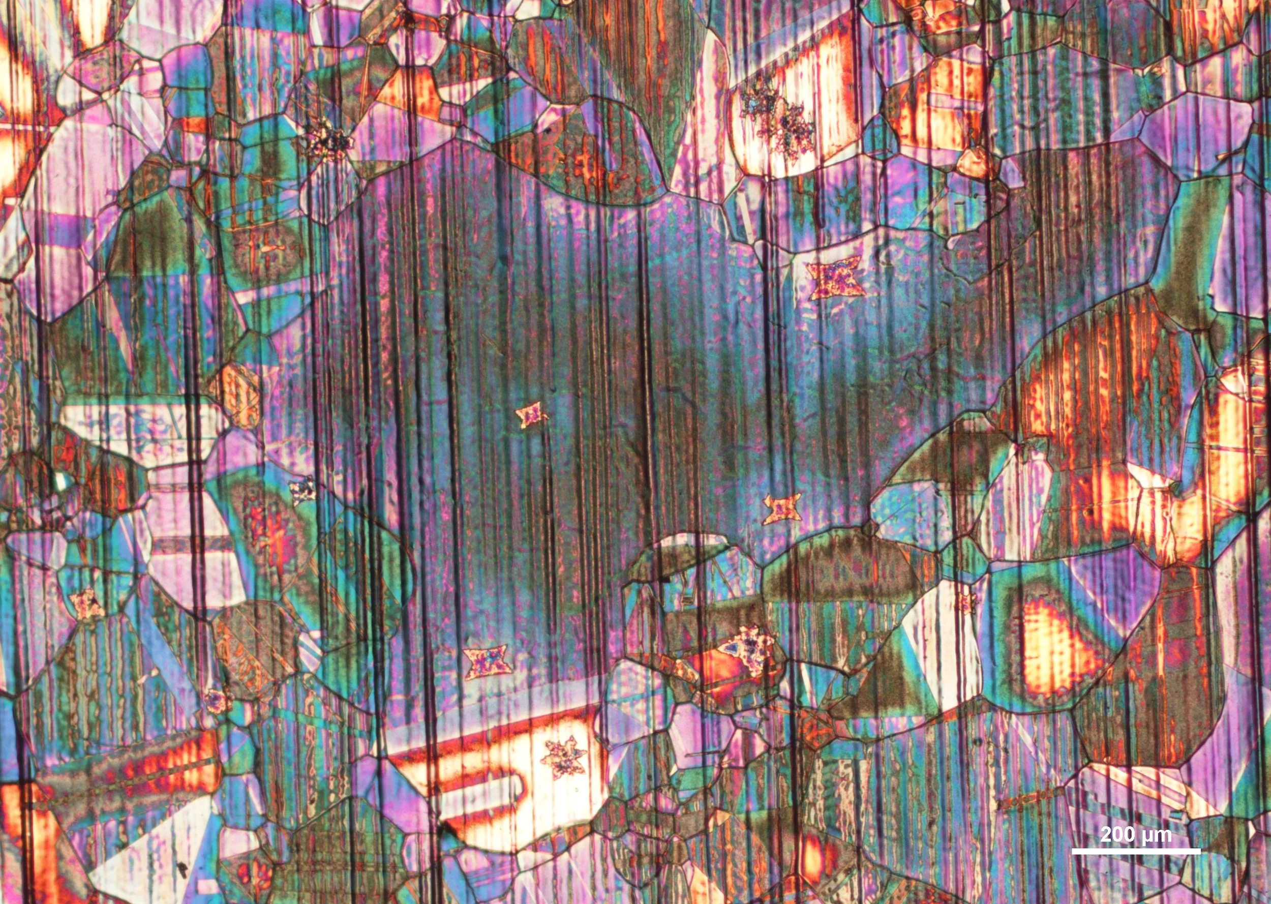
“Hidden Heart of Sustainability”
This optical microscope image shows the colourful microstructure of an aluminium alloy, made visible through special preparation and light interference. The ‘heart’ of the structure, formed thanks to recycled manganese, improves performance and expands its uses.
Captured at 1000X with a Zeiss Axio Observer 7 at the Czech Academy of Sciences.
Ondřej Ambrož, Postdoctoral Researcher at the Czech Academy of Sciences.
“A Heartbeat for a Sustainable World”
This SEM image shows the α-AlFeMnSi phase in an aluminium alloy, where manganese helps transform harmful needle-like structures into compact, often bizarre forms. This evolution reflects both the hidden beauty of materials and the promise of sustainable aluminium recycling.
Captured at Crytur with a Thermo Fisher Quattro SEM at 3000X magnification.
Ondřej Ambrož, Postdoctoral Researcher at the Czech Academy of Sciences.

“Perched in a Changing World”
A lone bird perches at dawn atop Serra de Montejunto, its silhouette framed by nature’s soft green hues. This image reminds us of the delicate balance of ecosystems and the urgent need to protect biodiversity amid ongoing environmental change.
João Martins, Molecular Biologist and Photographer passionate about scientific discovery and visual storytelling.

“Blue Pineapple”
At first glance, this vibrant, circular form may resemble a bacterial colony, but its folded hexagonal boron nitride (hBN) flakes, are transferred from a Cu foil onto a Si substrate. The folds, seen here in glowing blues and yellows, create a local strain that can subtly alter hBN’s wide bandgap (~5.9 eV), hinting at new possibilities for optoelectronic applications. A perfect fusion of art and science.
Elmahdi Amar, PhD student at INL - International Iberian Nanotechnology Laboratory and the Physics Center of Minho and Porto Universities (CF-UM-UP).

“A Cosmic Dance at the Microscale”
This confocal image captures chondrocyte cells in a hydrogel, with Sox9 expression glowing like stars in a galaxy. Beyond its beauty, it reflects progress in cell-based therapies for cartilage repair, offering hope for improved mobility and quality of life. Taken at i3S (University of Porto) using a Leica SP8 confocal microscope, it highlights how microscopic science drives medical innovation and eco-conscious solutions for the future.
Emine Kahraman, Postdoctoral Researcher at MIA Portugal in the Systems Cell Biology Group.
“Ammonia Borane Mini-boat”
Ammonia borane (NH₃BH₃) serves as a single-source precursor for hBN growth. Placed in a mini-boat inside a quartz tube within a hot-wall furnace, it decomposes under heat. A flow of argon and hydrogen carries the resulting boron and nitrogen radicals toward a copper foil, where hexagonal boron nitride forms.
Elmahdi Amar, PhD student at INL - International Iberian Nanotechnology Laboratory and the Physics Center of Minho and Porto Universities (CF-UM-UP).
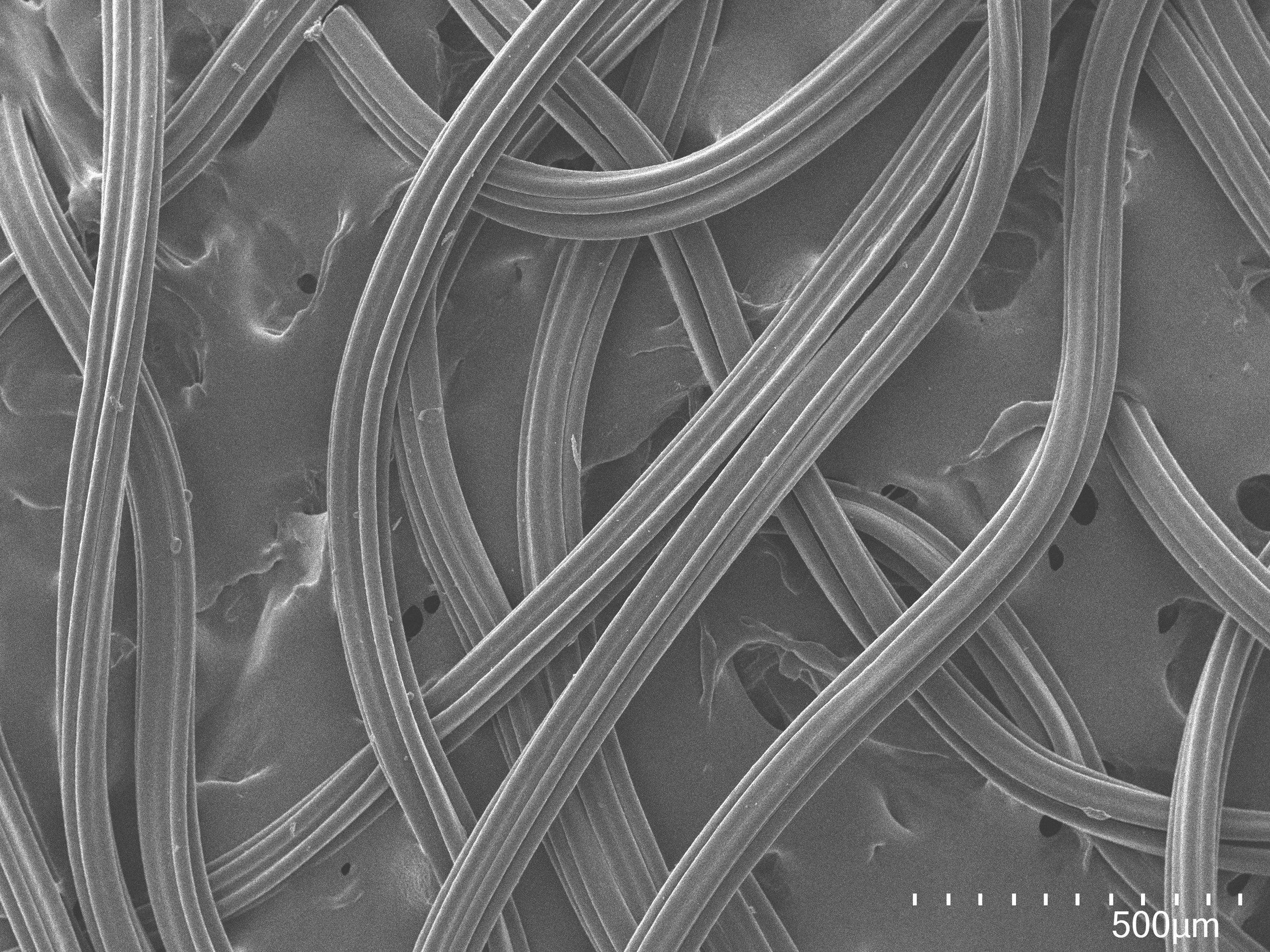
“Surface Morphology of Elastane Filament Under SEM”
This SEM image shows the microstructure of an elastane filament, known for its elasticity and resilience. The cylindrical fibre reveals surface details, including irregularities or effects from processing, offering insights into its mechanical behaviour. Captured at 2C2T – Centre for Textile Science and Technology, a leading research hub in textile materials and advanced microscopy.
Ana Catarina Silva works at ACS in the Lusitano project and contributes to innovation in textile recycling technologies, optimising separation and functionalisation processes for recycled fibres.

“Cosmografia del Minor Mondo”
This SEM image shows an elastane filament degraded by exposure to an organic solvent. The once smooth surface now appears rough, cracked, and brittle—evidence of polymer breakdown and loss of elasticity. Captured at 2C2T – Centre for Textile Science and Technology, this image highlights elastane’s chemical vulnerability and aids research on polymer resilience.
Temple Douglas is a Research Fellow in the Ultrafast Bio- and Nanophotonics group led by Jana Nieder at the International Iberian Nanotechnology Laboratory.
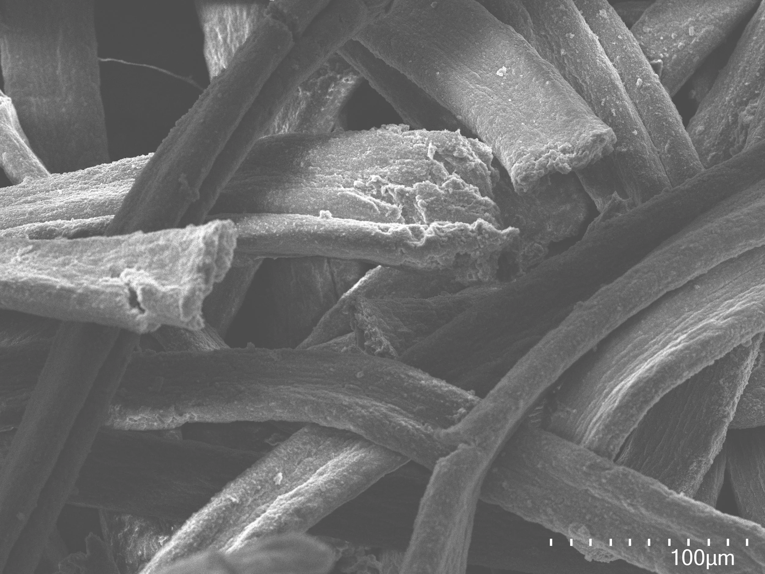
“SEM Image of Elastane Filament: Degradation Induced by an Organic Solvent”
This SEM image shows an elastane filament degraded by exposure to an organic solvent. The once smooth surface now appears rough, cracked, and brittle—evidence of polymer breakdown and loss of elasticity. Captured at 2C2T – Centre for Textile Science and Technology, this image highlights elastane’s chemical vulnerability and aids research on polymer resilience.
Ana Catarina Silva works at ACS in the Lusitano project and contributes to innovation in textile recycling technologies, optimising separation and functionalisation processes for recycled fibres.

“Elegant Lace”
The image demonstrates the silver chloride deposits electrochemically formed at the silver surface taken through a FEI Quanta 650 FEG scanning electron microscope (AEMIS Facility, INL) at 2,300x magnification. The scale bar shows 10 μm.
Olesia Dudik, Postdoctoral Researcher in the Espiña Research Group at the International Iberian Nanotechnology Laboratory (INL)-

“A Life in Motion: Modeling Disease in C. Elegans”
This image shows the life cycle of transgenic C. elegans expressing proteins linked to Machado-Joseph Disease, aiding research on neurodegeneration. As a simple model, C. elegans supports sustainable, efficient discovery. Captured with Confocal FV3000 (60X) at ICVS – University of Minho.
Daniela Vilasboas-Campos, Assistant Researcher, studying neural circuits and behaviour at the School of Medicine at the University of Minho in Braga, Portugal

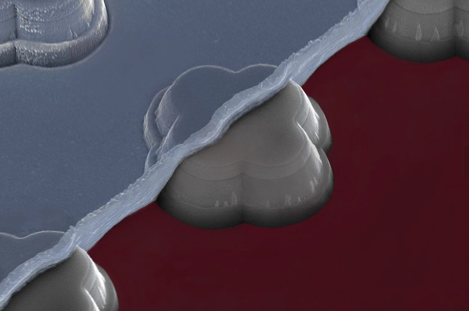
“A Hole in a Glass”
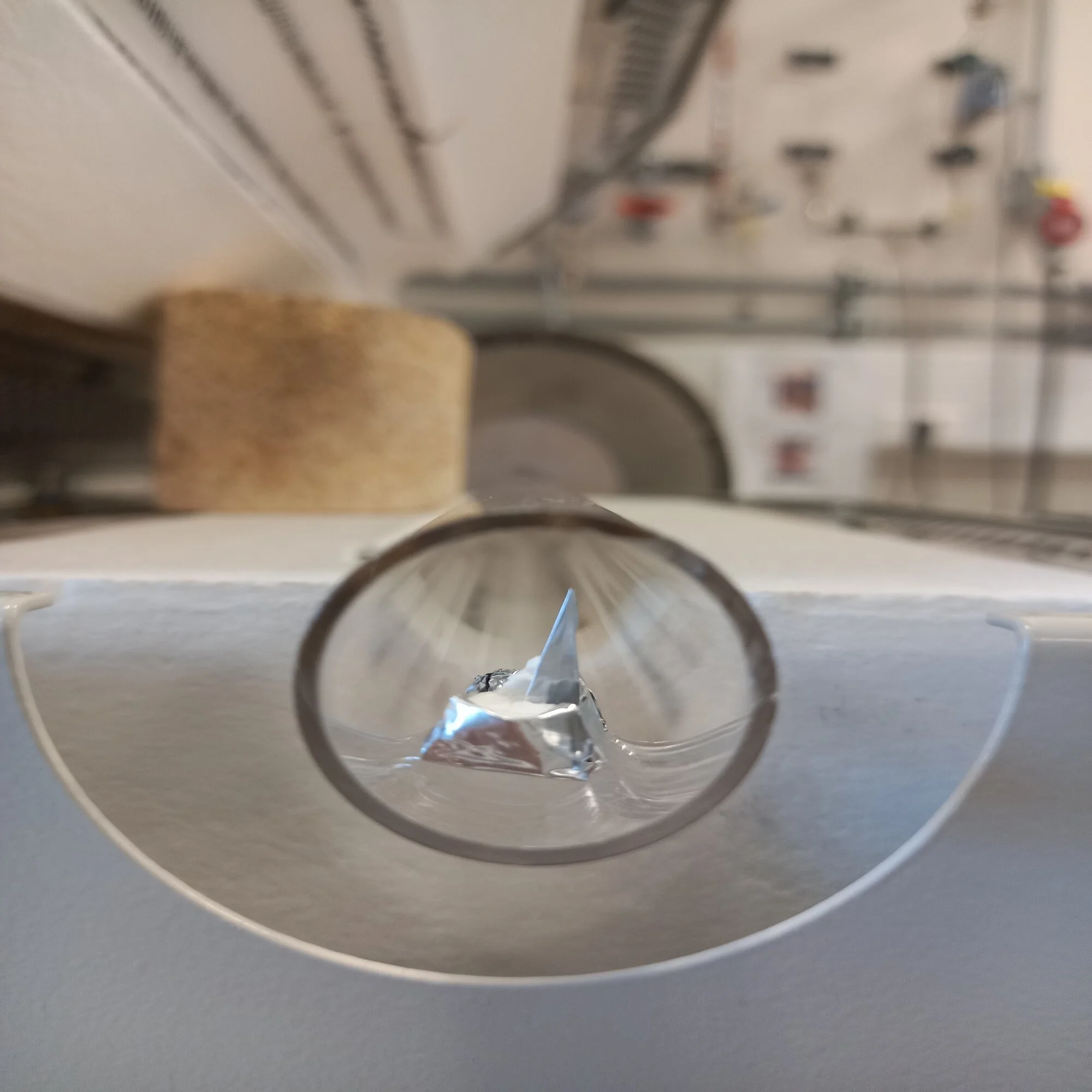
This photo is a cross-section of a sample that comprises the whole stack of a CIGS solar cell (ZnO: Al/ZnO/CdS/CIGS/Mo), around 3 um thick, deposited in 1mm glass. This is a great perspective on how small thin-film solar cells are. In this photo, it looks like the glass was hit by something, creating a hole, which resembles the craters in the moon.
Ana Margarida Moura, Research Assistant at the International Iberian Nanotechnology Laboratory (INL)-

“Illuminating Life: Tracking Cellular Responses in C. Elegans”
This image shows a C. elegans hlh-30 reporter strain, used to study stress responses and metabolism. The left panel is a brightfield view; the right shows fluorescent HLH-30, a transcription factor linked to autophagy and longevity. Genetically engineered C. elegans enable ethical, sustainable research into key biological processes. Captured with Confocal FV3000 (60X) at ICVS – University of Minho.
Daniela Vilasboas-Campos, Assistant Researcher, studying neural circuits and behaviour at the School of Medicine at the University of Minho in Braga, Portugal
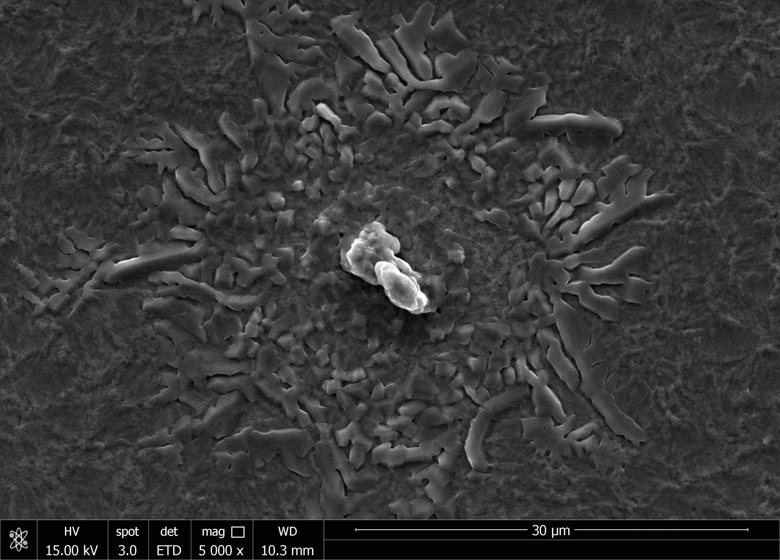
“Oxygen Explosion”
This photo is the morphology of the surface of air-oxidized Mo. MoOx is an intralayer of elemental Se solar cells. This is a defect in the film that looks like an explosion.
Ana Margarida Moura, Research Assistant at the International Iberian Nanotechnology Laboratory (INL)-
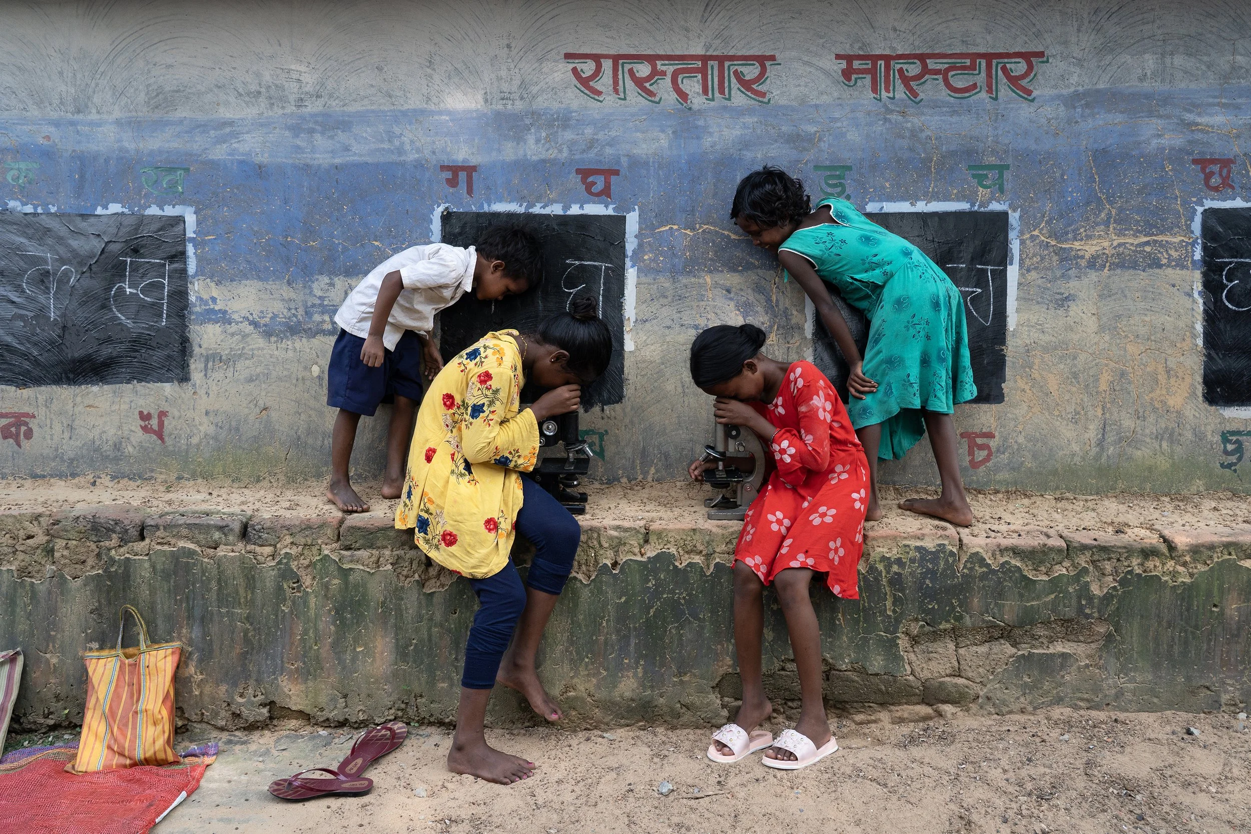
“Microscopic Mythbusters”
In remote West Bengal, India, tribal children explore the microscopic world through the Teacher of the Streets program. This initiative brings education to those unable to attend school, combating harmful superstitions with science and sparking curiosity through tools like microscopes.
Sudip Maiti is an Independent Photojournalist based in Kolkata, India.

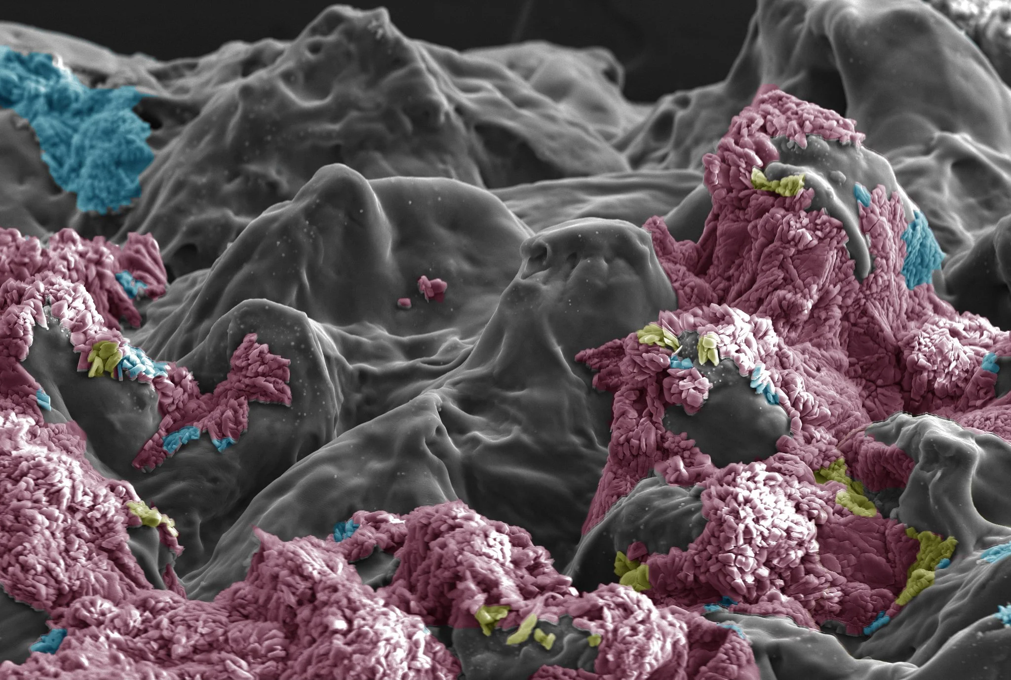
“Love for the Planet: A Bacterium’s Heartfelt Glow”
In the spirit of Science for a Sustainable Future, Vibrio fischeri ‘wears its heart’—a symbol of how research drives environmental and societal resilience. This SEM image, taken at INL during my PhD, features V. fischeri used as a toxicity biosensor, as detailed in my publication: https://doi.org/10.3390/chemosensors9100283.
Ana Rita Silva, a Biomedical Engineer currently Invited Auxiliary Researcher at the Centre of Biological Engineering (CEB) at the University of Minho.
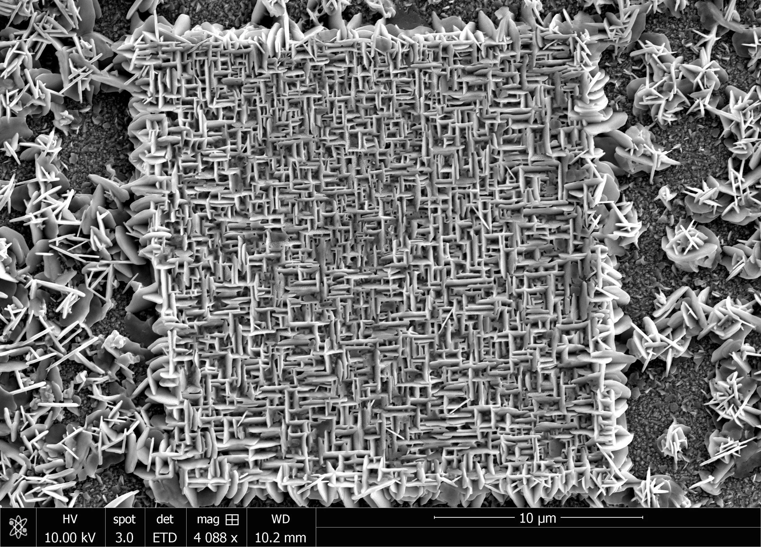
“Microlabyrinth”
This SEM image from INL’s cleanroom shows a 10×10 microLED array, with 100 precisely engineered LEDs and intricate top contacts forming a maze-like, 3D visual effect. The 900 µm square structure showcases advanced microfabrication, suited for high-resolution displays, visible light communication, and optogenetics requiring precise light control.
Nuaman Moideen Kutty is a dedicated Research Assistant at the International Iberian Nanotechnology Laboratory(INL), specialising in photonics and laser physics.
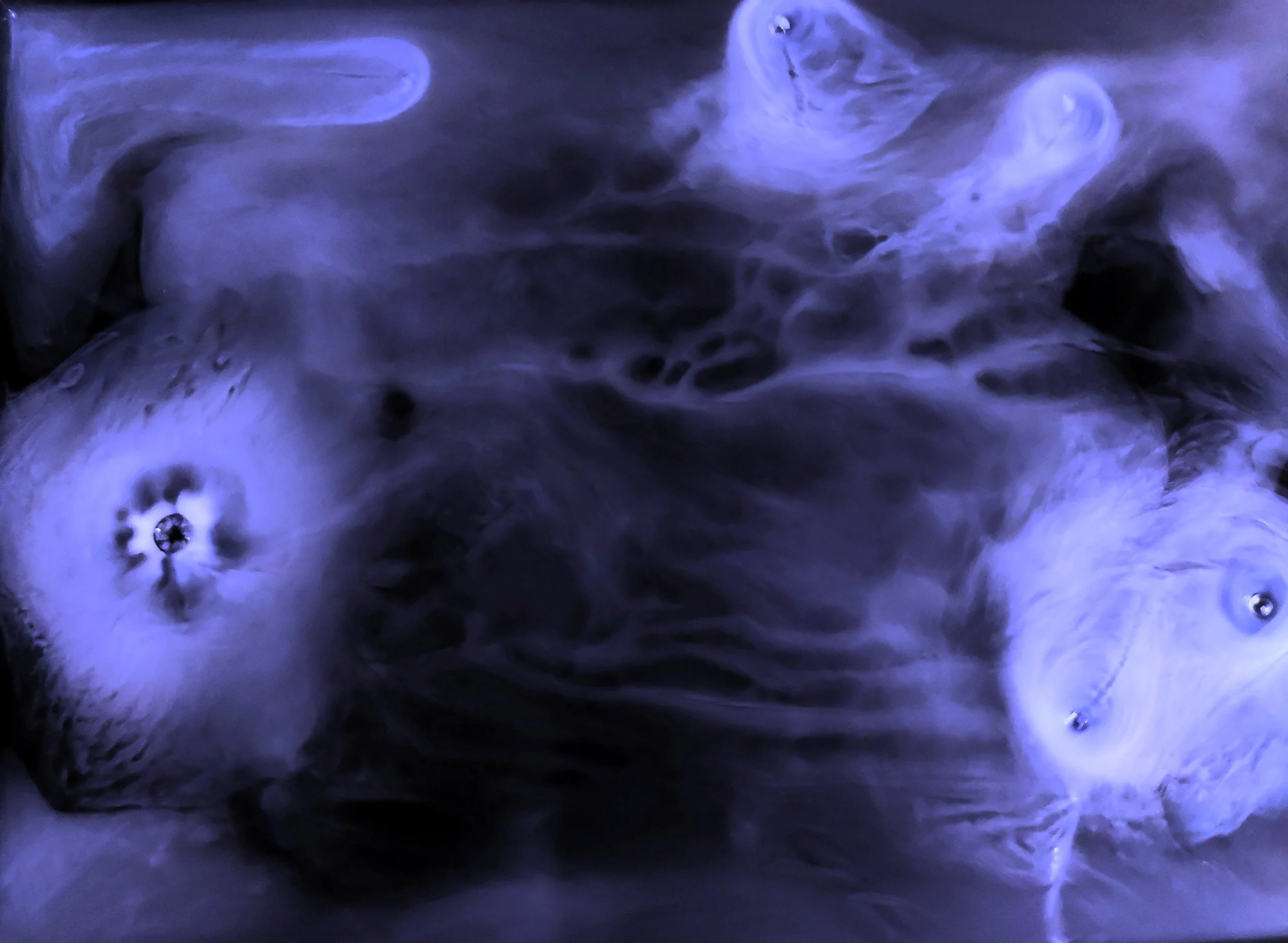
“Subzero Nebulae”
This image shows liquid nitrogen droplets skimming over alcohol due to the Leidenfrost effect. The extreme temperature difference causes nitrogen to boil instantly, forming a vapour layer that lets the droplets glide with minimal friction.
Daniel Abella López, Pre-Doctoral Researcher at CiQUS-Singular Center for Research in Biological Chemistry and Molecular Materials.

“Maze of Light”
This SEM image from INL’s cleanroom shows a 5×5 microLED array (400 µm square), with intricate top contacts creating a maze-like, 3D visual effect. The design exemplifies precision microfabrication and supports advanced applications in high-res displays, visible light communication, and optogenetics requiring precise light control.
Nuaman Moideen Kutty is a dedicated Research Assistant at the International Iberian Nanotechnology Laboratory(INL), specialising in photonics and laser physics.
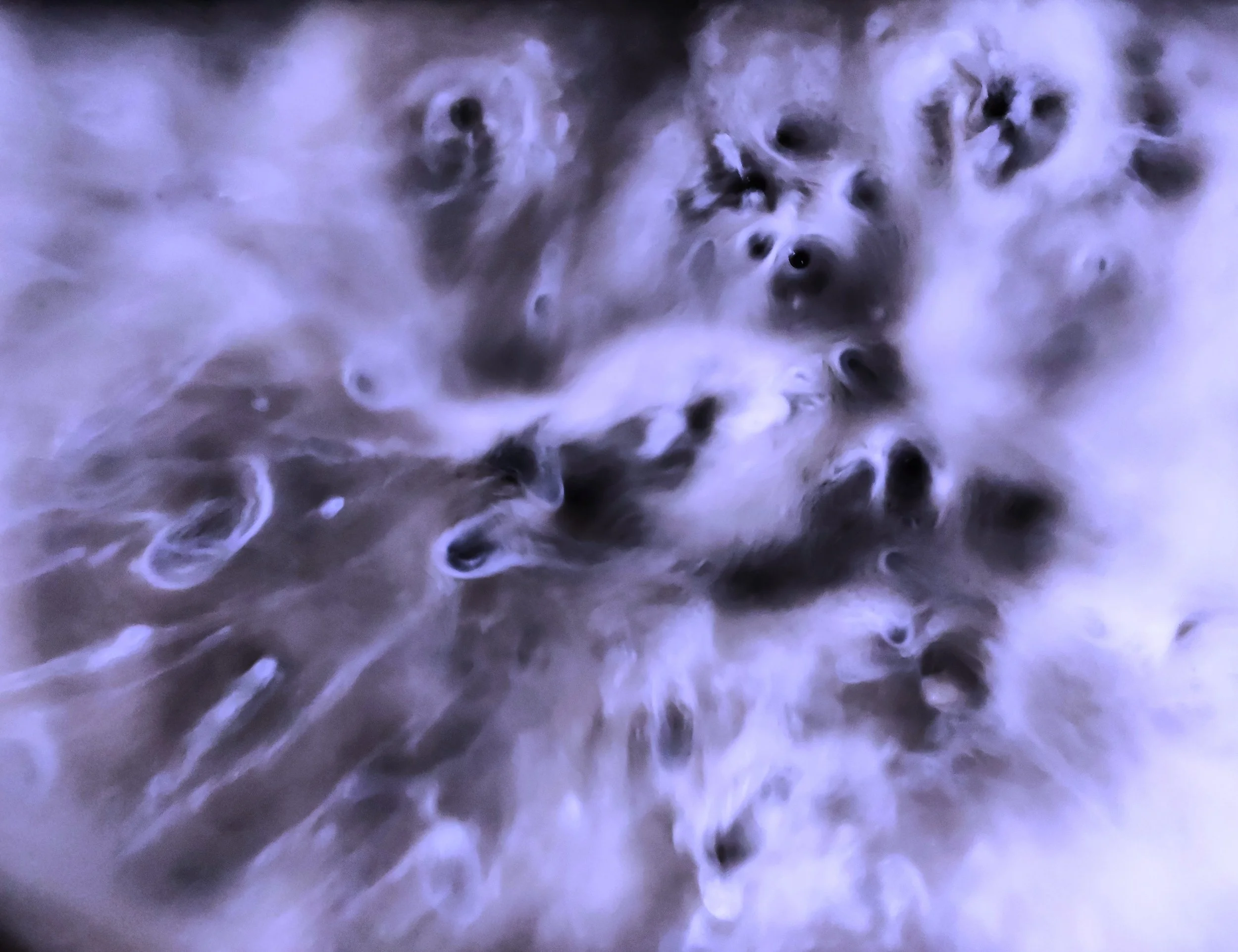
“Universe in Formation”
Resembling a starry universe, this image shows liquid nitrogen droplets on alcohol, demonstrating the Leidenfrost effect. The extreme temperature difference causes nitrogen to boil instantly, forming a vapour layer that lets droplets glide freely with minimal friction."
Daniel Abella López, Pre-Doctoral Researcher at CiQUS-Singular Center for Research in Biological Chemistry and Molecular Materials.
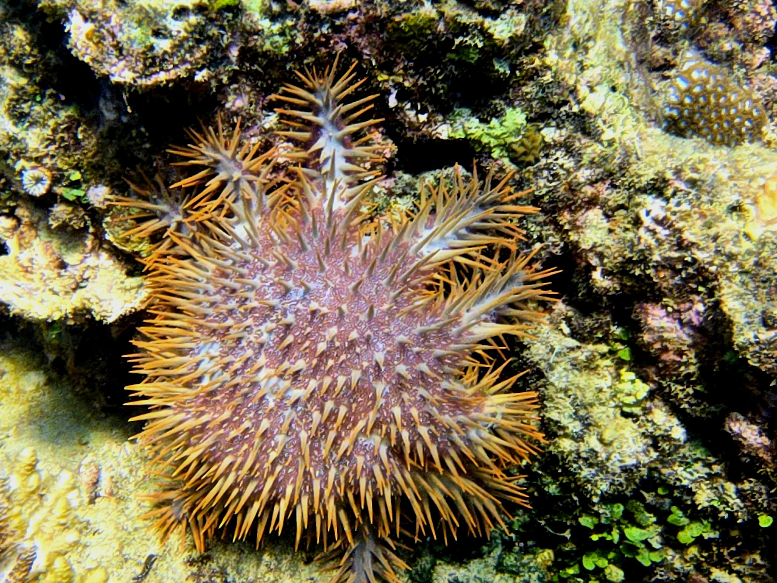
“The Venomous Starfish”
Monitoring the population of the venomous Crown-of-Thorns Starfish (COTS) is one of the most important tasks of MPA managers. Outbreaks of COTS are responsible for the extensive loss of reef-building corals. Hence, an early detection of COTS infestation means early interventions.
Jane Domingo-Dela Cruz, a wife and a mom. Works in the Department of Environment and Natural Resources under the Conservation and Development Section, Philippines
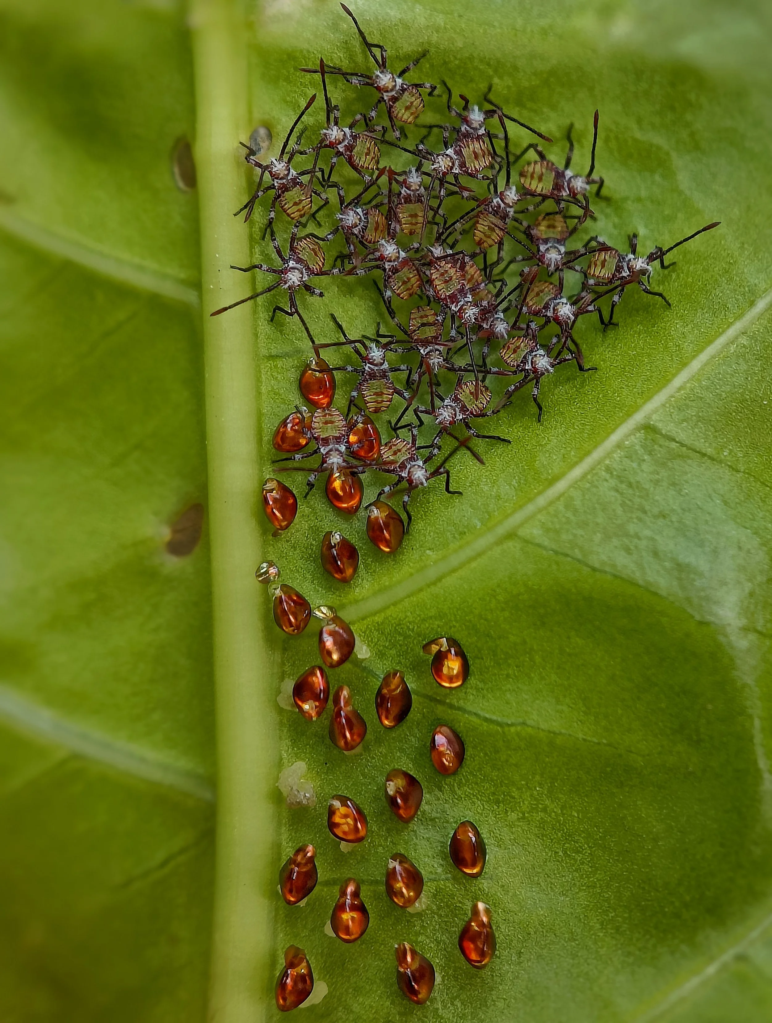
“Nymphs and Nature: A Close-Up Journey”
This macro photo captures newly hatched Acanthocoris scaber nymphs nestled under leaf foliage. Their soft, translucent bodies and clustered behaviour reflect a survival strategy that offers protection and resource access. These tiny insects play a vital role in biodiversity, plant health, and ecological balance.
Sritam Kumar Sethy, Nature Enthusiast and Wildlife Photographer based out of Odisha, India.

“Mastering the Art of Camouflage”
Eat or be eaten—that's nature's rule. This image shows an Asian grass frog (or Boei's wart frog) camouflaged against tree bark, using crypsis to avoid predators and ambush prey like insects and worms. Its bark-like skin aids survival through stealth, making it both predator and prey in the delicate balance of the ecosystem.
Sritam Kumar Sethy, Nature Enthusiast and Wildlife Photographer based out of Odisha, India.
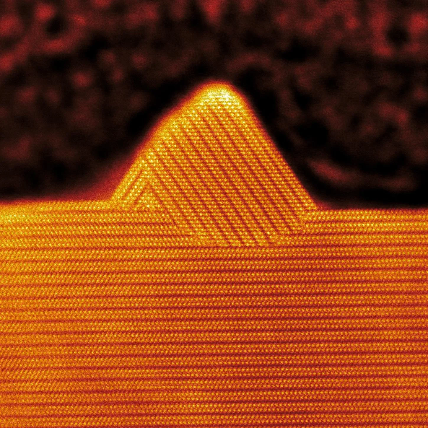
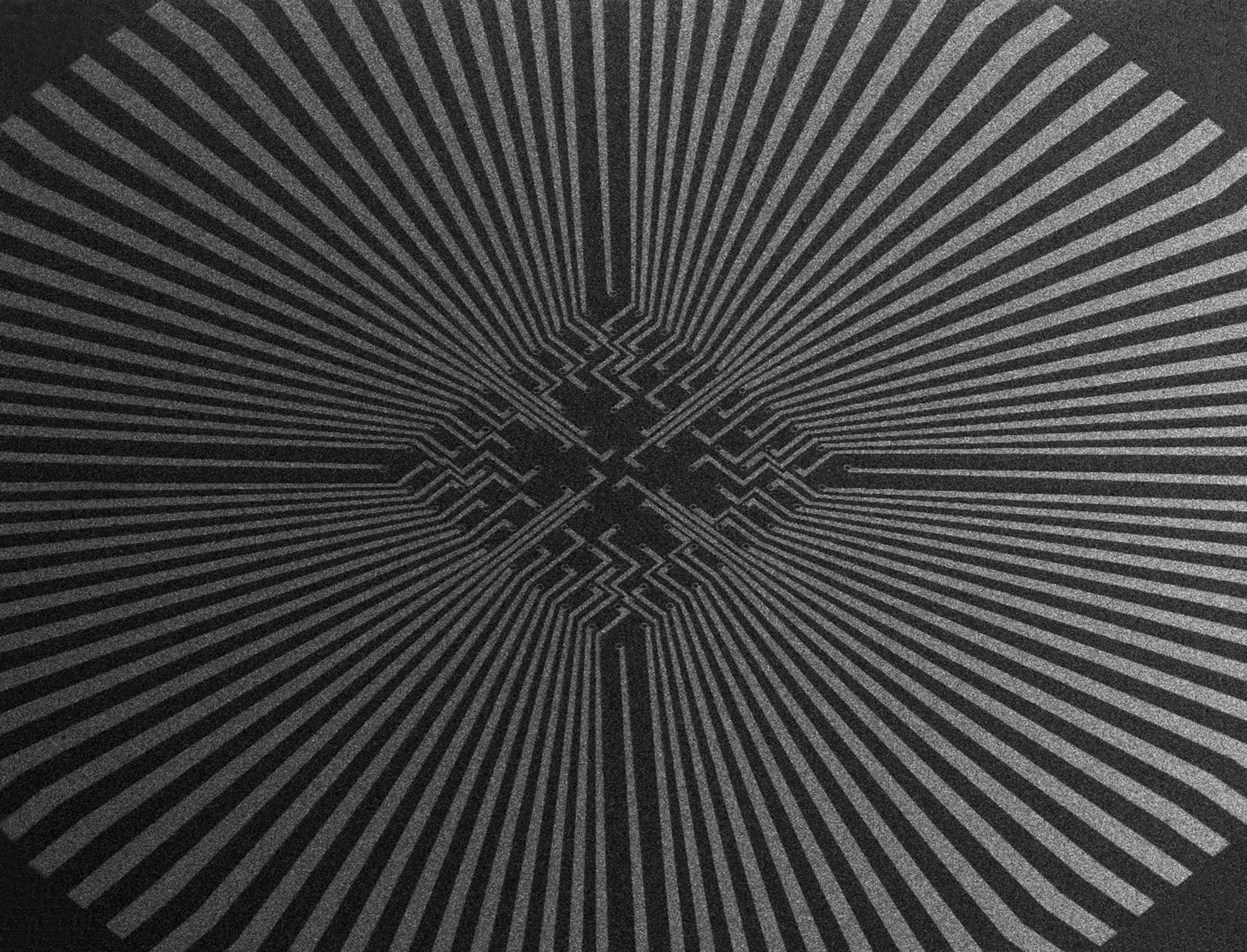
“Nanoscale Mountains”
This HR-STEM image reveals the nanoscale 'mountain-like' surface of a Cu-doped Bi₂Se₃ thin film, shaped by complex atomic processes. As a topological material, Bi₂Se₃ offers potential for energy-efficient electronics and thermoelectrics. Copper doping enhances its properties, supporting advances in sustainable tech and quantum computing.
James Caleb Peters, a researcher exploring the fascinating world of quantum materials, focuses on understanding the structural and electronic transformations that occur at the atomic scale.
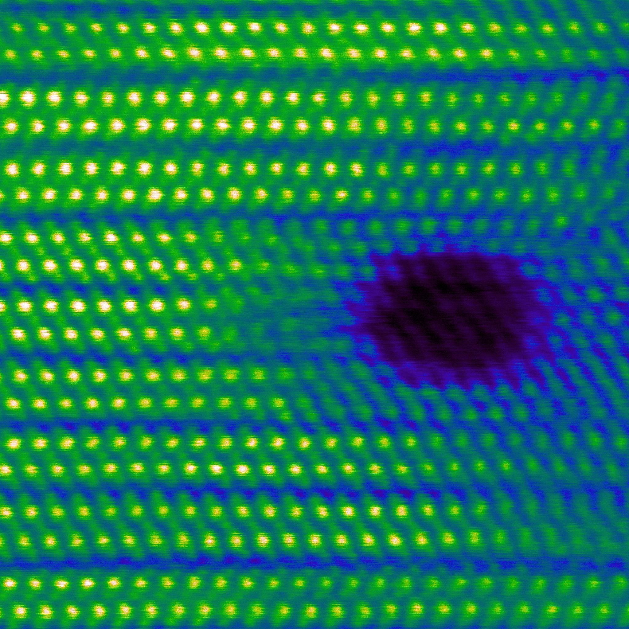
“Nano Scar: The Mark of the Electron Beam”
This HR-STEM image of a Cu-doped Bi₂Se₃ thin film shows a 'nano scar',an artifact from electron beam interaction. While unintentional, it reveals the fine balance between material stability and imaging precision. Understanding such effects is vital for advancing synthesis and characterization of quantum materials for sustainable, energy-efficient technologies.
Captured at INL using an FEI Titan ChemiSTEM.
James Caleb Peters, a researcher exploring the fascinating world of quantum materials, focuses on understanding the structural and electronic transformations that occur at the atomic scale.

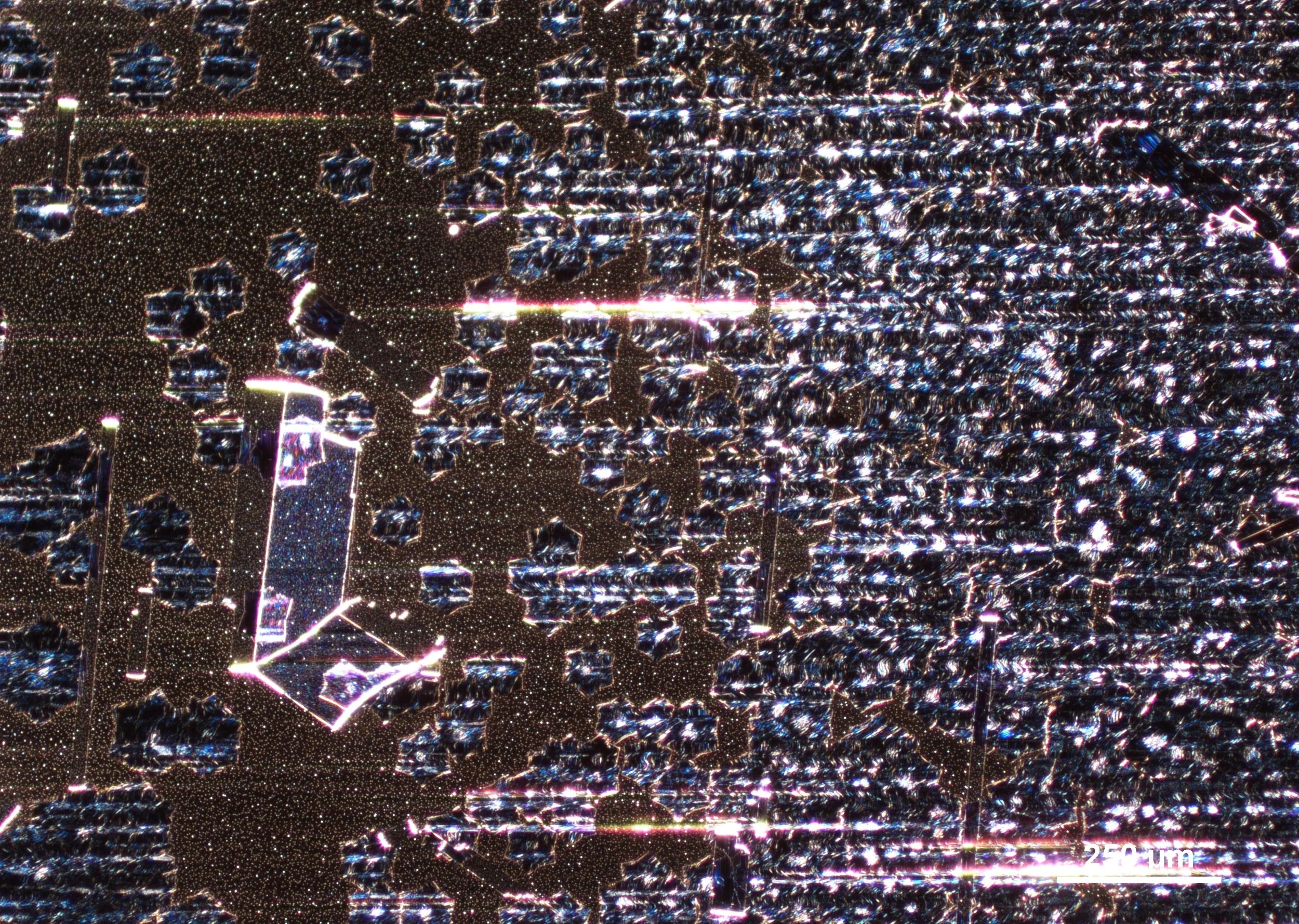
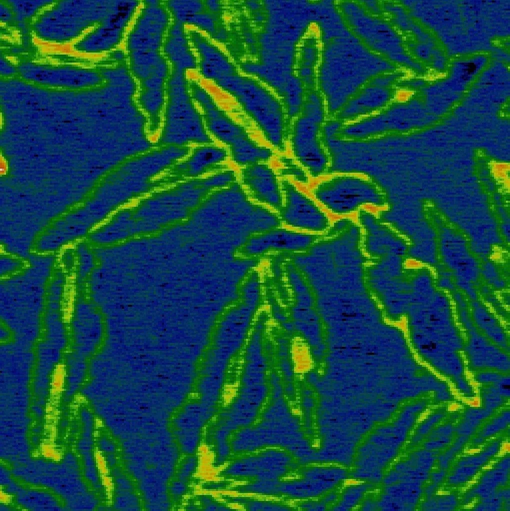
“Crystal Leaves”
Captured at 100x magnification, this image shows a nucleation region in a crystalline film made from a chemical mixture. Though transparent under normal light, crossed polarisers reveal a leafy, botanical-like pattern formed by self-assembled crystal grains. It highlights the hidden beauty and order emerging from molecular self-organization.
Captured at The Art of Science Photography studio.
Karl Gaff, with a background in physics and expertise in advanced imaging techniques, captures the hidden beauty of crystals using polarised light microscopy, blending science and art.
“Nano-Raspberries: A Berry Good Solution for the Future?”
This SEM image shows raspberry-like nanostructures used in anti-graffiti coatings. Their superhydrophobic properties prevent paint adhesion, reducing chemical cleaning and supporting sustainable, cleaner urban spaces.
Juliana Sousa, Researcher at INL with expertise in Inorganic Chemistry and Materials Science, specializing in the synthesis and functionalization of inorganic materials
“Cs₂AgBiBr₆ Halide Double Perovskite Porous Single Crystals”
This SEM image shows porous single crystals of Cs₂AgBiBr₆ perovskites formed via infrared spin coating. Oriented platelets combine into porous clusters, with pores mainly between the crystal layers. Captured at the Nanostructured Materials group at INL.
Sandeep Maurya, Postdoctoral Researcher (Postdoc) at Nanostructure Materials group at the International Iberian Nanotechnology Laboratory (INL),
“Rose Lens”
Created using a Diffractive Optical Element (DOE), this image combines red, green, and blue wavelength data into an RGB composition. The rose window-like patterns reveal the DOE’s diffractive structure, captured via a custom microscope setup.
Manuel Rodrigues, Postdoctoral Researcher, Integrated Micro and Nanotechnologies Research Group at the International Iberian Nanotechnology Laboratory (INL),
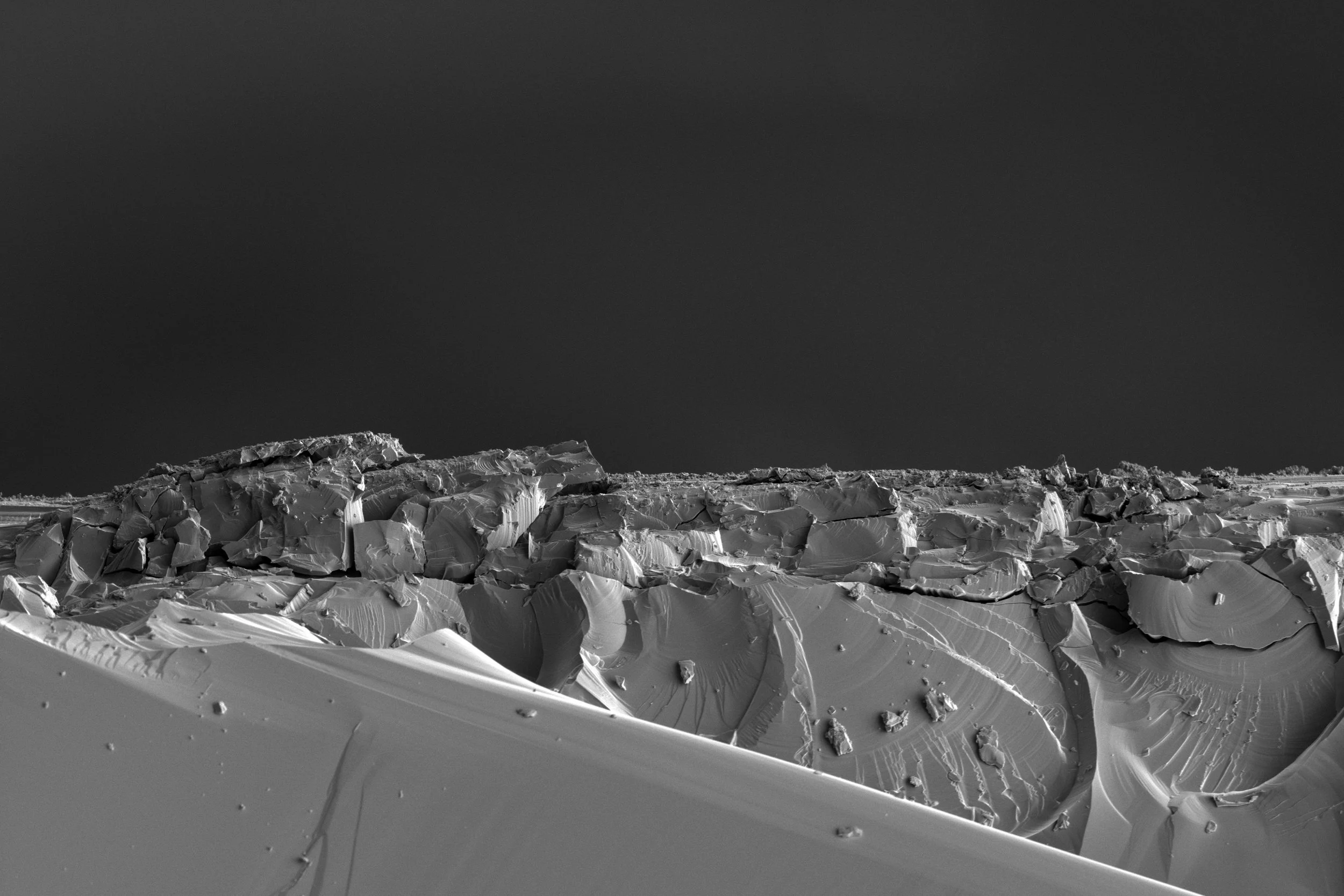
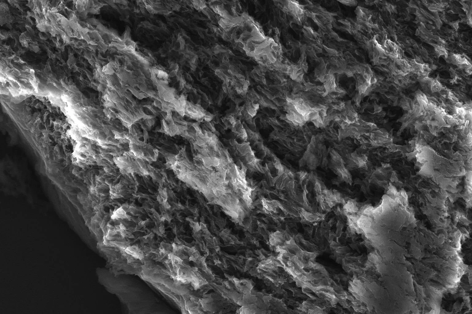
“Silicon Moon”
This SEM image shows the rough fracture edge of a small silicon sample, revealing a surface that strikingly resembles the moon’s landscape. A reminder, as Blake wrote, that we can 'see a world in a grain of sand.
Manuel Rodrigues, Postdoctoral Researcher, Integrated Micro and Nanotechnologies Research Group at the International Iberian Nanotechnology Laboratory (INL),
“The Lonely Star”
Darkfield image of CVD-grown graphene on copper foil. Large flakes merged into a continuous film with dendritic, star-like patterns. A single isolated flake, unable to join its neighbours, creates a gap in the 'graphene space.' Water-soluble polymers enabled eco-friendly dry transfer.
Tiago Pereira, Research Engineer in the 2D Material and Devices group at the International Iberian Nanotechnology Laboratory (INL).
“Fragmented”
Darkfield image of CVD-grown graphene on copper foil. Hexagonal flakes align and merge into a single-crystal film. A copper twin-grain forms a dark-blue rectangle—like a doorway into another graphene universe.
Tiago Pereira, Research Engineer in the 2D Material and Devices group at the International Iberian Nanotechnology Laboratory (INL).

“It’s a plane? Is it a bird? No, it’s Superhydrophobic Coating!”
This image shows a glass slide coated with a highly hydrophobic, fluorine-free coating developed in my research at INL. Such coatings repel water and have wide applications—from waterproof clothing to corrosion protection—offering both industrial and environmental benefits.
Diana Alves, Research Fellow of the Nanochemistry group at INL - International Iberian Nanotechnology Laboratory. Her research is focused on developing superhydrophobic coatings.

“Sharp Science for a Future On-point"
SEM image of needle-like zinc oxide synthesized for hydrophobic coatings. Surface roughness and controlled morphology enhance water-repellent properties. Captured at INL with a Quanta FEG 250 SEM at 5000X magnification.
Diana Alves, Research Fellow of the Nanochemistry group at INL - International Iberian Nanotechnology Laboratory. Her research is focused on developing superhydrophobic coatings.
“Crystal Starships Invasion”
In a galaxy far away, thousands of crystal starships are propelled through deep space, surrounding and subsiding the enemy. Photograph of nanocrystals on a copper foil taken by scanning electron microscopy.
Mafalda Abrantes, Physics Engineer of the Alpuim Research Group at INL - International Iberian Nanotechnology Laboratory.
"Graphene Stars on a Copper Desert”
Graphene stars on a copper desert. Photography taken with an optical microscope.
Mafalda Abrantes, Physics Engineer of the Alpuim Research Group at INL - International Iberian Nanotechnology Laboratory.
“Charged Algae”
This KPFM image shows biogenic selenium-based nanosystems (4×4 µm scan) developed for cancer research. Our work focuses on zero-valent selenium, known for its strong anticancer potential.
Captured using a SmartSPM (Horiba Scientific).
Marina Temiryazeva, Horiba Scientific, France

“Magnetic Snowflakes”
MFM image of CoPt/YIG multilayer structures (24×24 µm), key materials in spintronics. These delicate magnetic 'snowflakes' highlight both technological progress and the fragility of our planet.
Captured using a SmartSPM (Horiba Scientific).
Marina Temiryazeva, Horiba Scientific, France
“Brain Gold”
Brain Gold is an image taken with Scanning Electron Microscopy and is a composite of COF with gold.
Miguel Chaves, a Chemist with a Master’s degree in Chemistry currently works as a Research Assistant at the International Iberian Nanotechnology Laboratory (INL).
“Flymer”
Flymer is an image taken with Scanning Electron Microscopy and is a Covalent organic polymer dry synthesised.
Miguel Chaves, a Chemist with a Master’s degree in Chemistry currently works as a Research Assistant at the International Iberian Nanotechnology Laboratory (INL).
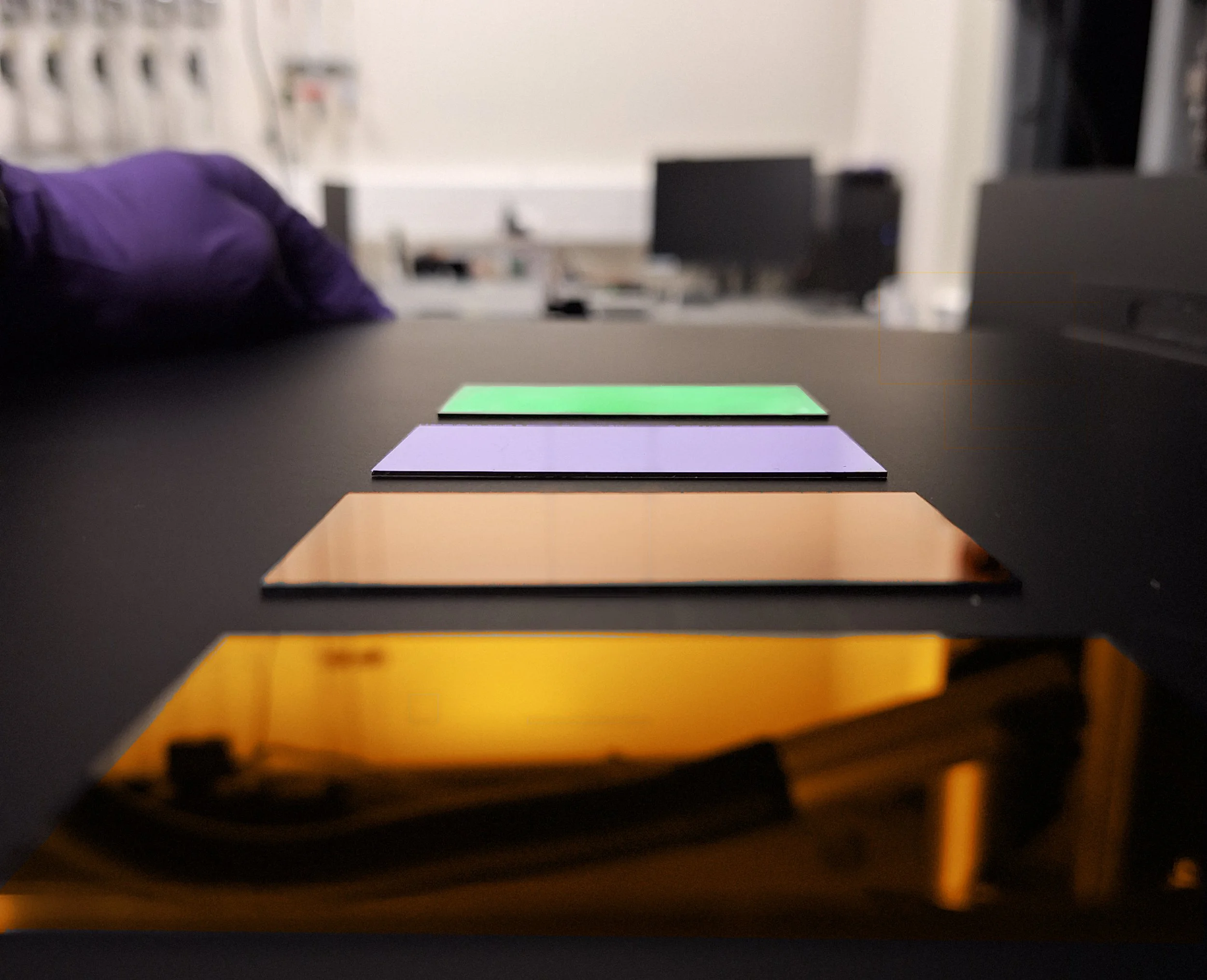
“TiO₂ Reflections: Towards a Greener Future”
This photo shows TiO₂ thin films made with different oxidising agents to create amorphous layers for UV applications. As a non-toxic, eco-friendly material, TiO₂ supports sustainable tech. The samples, placed on a Thorlabs anodised screen, reflect vivid colours—symbolising progress toward a greener future. The greenest, most recent sample is featured last.
Diogo Poeta, Research Assistant working on nanophotonics and sustainable materials at the Diéguez Research Group at the International Iberian Nanotechnology Laboratory (INL).
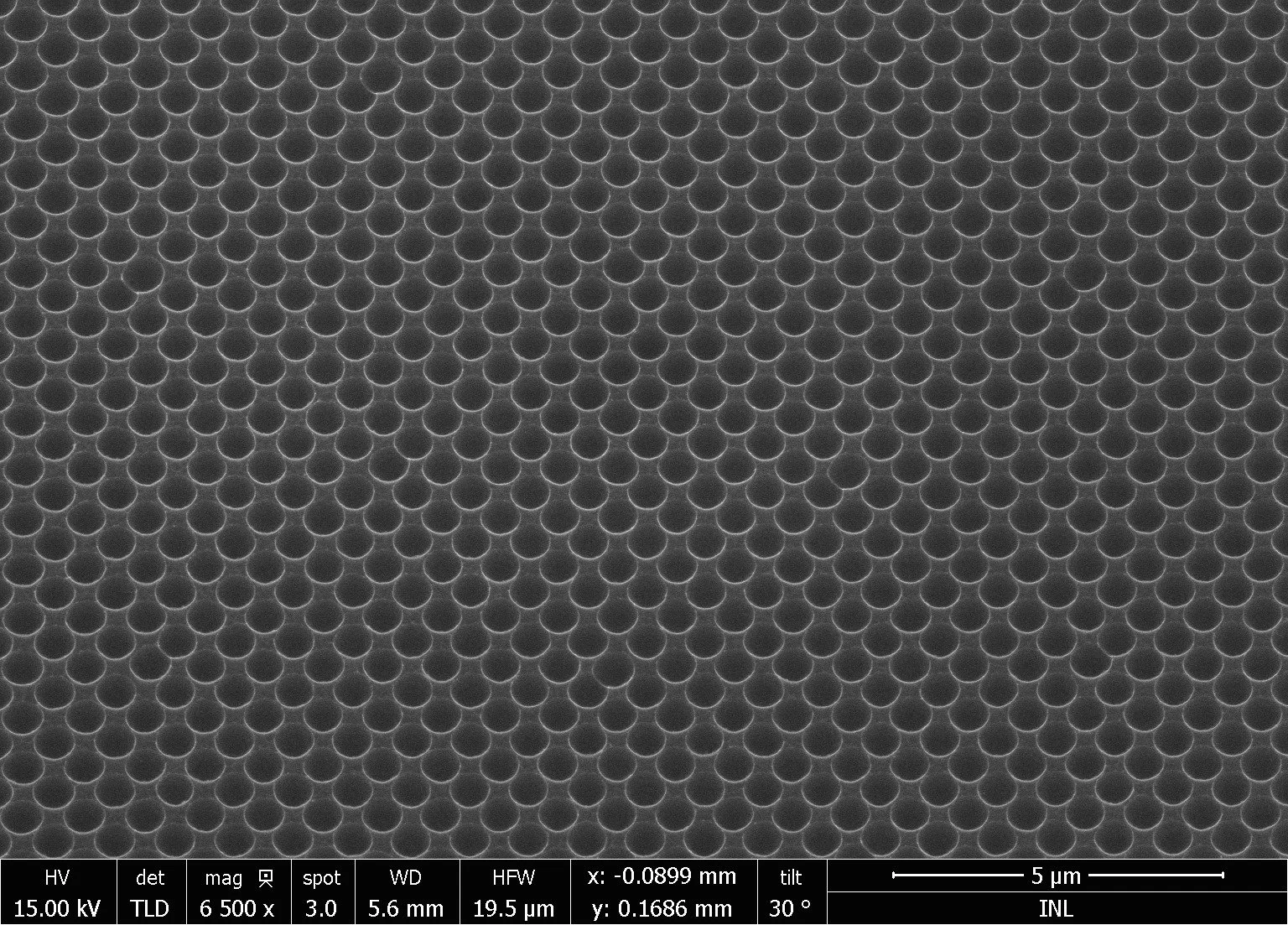
“Dragon Scales”
SEM image of 500 nm hemispherical concavities patterned on a silicon wafer to enhance solar cell efficiency. Rotated 90°, the array resembles dragon or fish scales. These nanostructures help boost light-to-power conversion.
André Violas, PhD Candidate at Aveiro University and the International Iberian Nanotechnology Laboratory (INL) in Portugal, in collaboration with Uppsala University in Sweden, and his research is focused on bifacial Cu(In, Ga)Se₂ (CIGS) solar cells.

“Yummy Rainbow Wafer”
A photo taken near the INL garden shows a 20 cm Si wafer with patterned SiO₂ squares. These patterns act like prisms, splitting sunlight into colours—an effect also useful for enhancing solar cell performance.
André Violas, PhD Candidate at Aveiro University and the International Iberian Nanotechnology Laboratory (INL) in Portugal, in collaboration with Uppsala University in Sweden, and his research is focused on bifacial Cu(In, Ga)Se₂ (CIGS) solar cells.
Have a question? We’re here to help!
If you have any doubts about the Photo Contest 2025, feel free to reach out using this form. Whether it's about participation, rules, deadlines, or submissions—we’re happy to assist!
Fill in your details and message below, and we’ll get back to you as soon as possible.
Unforgettable contest!
The Photo Contest 2025 Team
