
Exploring Science, Technology and Art
#ERN2022 Photo Competition Gallery
#ERN2022 Photo Competition celebrates science, technology and art in the most creative way. The most eclectic mix of ideas, disciplines and perspectives creates the most far-reaching conversations, in-depth debates, and inspiring developments.
This was an open competition and everyone was encouraged to submit up to two original photographs within the following categories::
→ General: Other images related to the scientific world.
→ Nano/micro: Images obtained through microscopes.
Your talent and unique visualisations were honoured and celebrated.
→ General Category
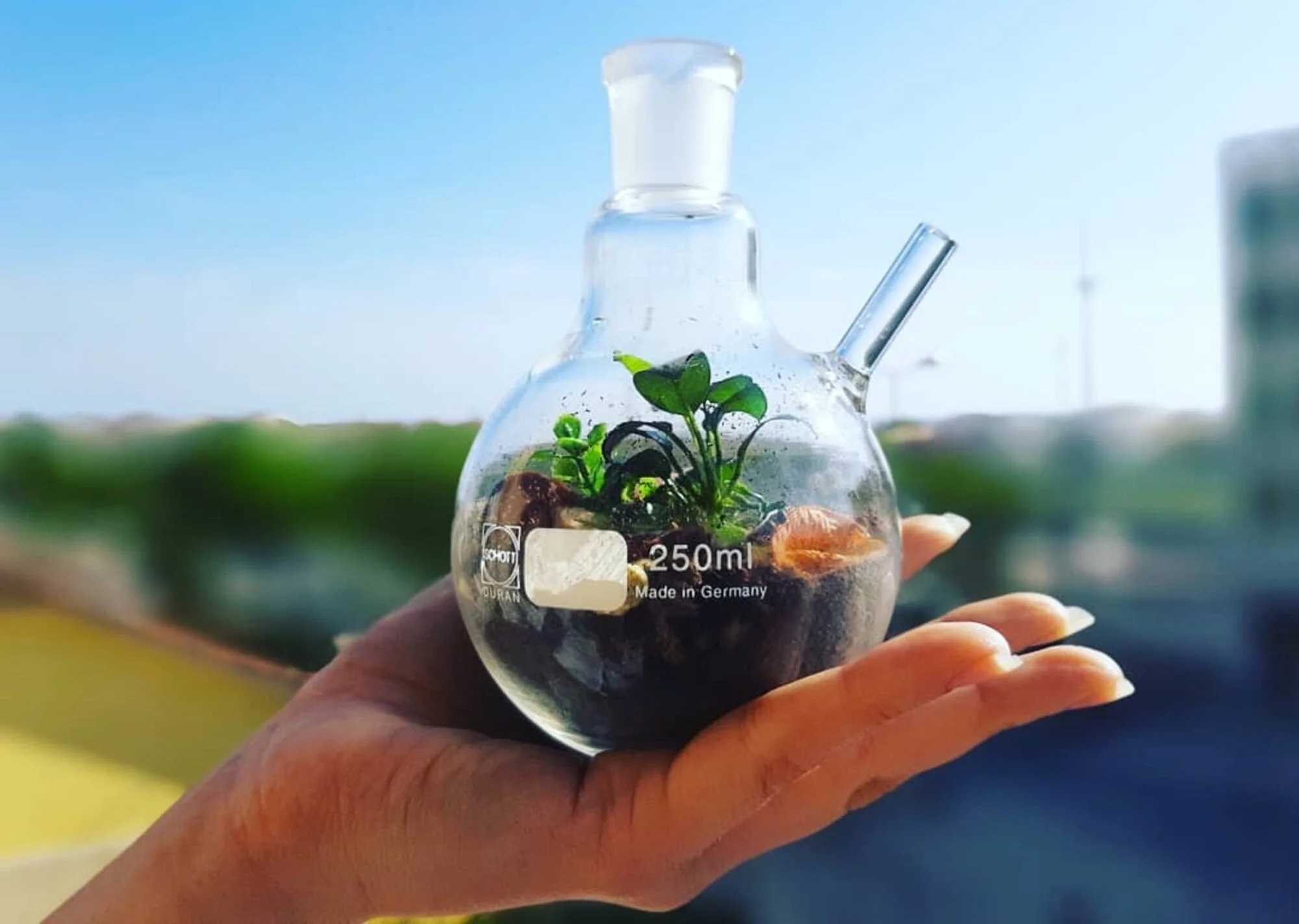

CARINA CRUCHO
Sustainability in an RB
This photo illustrates how science can help to create a sustainable world and raise awareness of the need to connect nature and science to reduce the harmful impact of humanity on the world. The photo was taken during the pandemic at home in lockdown to celebrate earth day on social networks.


MIGUEL XAVIER
The Scientist Emoji
Everyone knows that scientists spend their life boasting a white lab coat in a laboratory setting and playing with bubbling and fuming green fluorescent solutions.


HéLDER FONSECA
MirrorTech - see you on the white side
A productive silicon wafer was used to reflect the cleanroom's daily operations and its environment.
This photo was taken inside the INL Cleanroom.


CARLA GASPAR
Iminente #4
Iminente is a series of photos of a set of objects photographed in a stereoscopic phantogram, in anaglyph. They should be seen at 45 degrees (with red and cyan filter glasses*) horizontally, so that the projected figure appears in 3D, vertically suspended in the air. The objects represent a state of suspension, frozen in time and space, opposing the gravity of their weight. They can be penetrated in order to invade their inner space, or sustained by the observer’s hand or another object to prevent their fall. The piece enables interaction and duality between the two bodies, the virtual and the physical. Fragility and Resistance.
This photograph depicts several test tubes from a Physics and Chemistry Secondary School Lab in Braga.

CARLA GASPAR
Iminente #5
Iminente is a series of photos of a set of objects photographed in a stereoscopic phantogram, in anaglyph. They should be seen at 45 degrees (with red and cyan filter glasses*) horizontally, so that the projected figure appears in 3D, vertically suspended in the air. The objects represent a state of suspension, frozen in time and space, opposing the gravity of their weight. They can be penetrated in order to invade their inner space, or sustained by the observer’s hand or another object to prevent their fall. The piece enables interaction and duality between the two bodies, the virtual and the physical. Fragility and Resistance.
This photograph depicts several test tubes from a Physics and Chemistry Secondary School Lab in Braga.

HÉLDER FONSECA
You shall not pass!
It is from a Silicon wafer where some coatings delaminated caused by mechanical stress. The perpendicular shape acts as a physical stopper for the detachment.
This photo was taken with a standard camera.

JOÃO AVO
Layered Light
This photo depicts three different luminescent compounds that were developed for application in OLEDs in order to increase their efficiency. The compounds were dissolved in three non-miscible solvents to achieve the separation of colour. Each colour represents a primary colour of light, and their combination is used to achieve white light.

JOÃO MARTINHO MOURA
Sci-fi Miners. The exhibition was in Seoul, South Korea 2019
Large-scale exhibition at Art Center Nabi, in Seoul.
Sci-fi Miners: an artistic exploration of how, with the help of scientific advances in nanotechnology and artificial intelligence, a new generation of nano-clusters are replacing critical natural resources becoming rare on planet earth. Work developed at INL – International Iberian Nanotechnology Laboratory, and Aalto University. After the audiovisual and VR performance event at INL last April, a new exhibition with multiple large façade displays and virtual reality experience at Art Center Nabi, in South Korea (“Confluence Point”, Sci-fi Mines, João Martinho Moura, 2019). For the VR experience, please consult Art Center Nabi for availability.
Work designed by João Martinho Moura with the support of the STARTS Residencies as part of the STARTS program of the European Commission, based on the technological elements of the CritCat Project.

JOÃO MARTINS
A Mussel!
A Macro photo of a mussel taken in its natural habitat, during the low tide of Magoito Beach, Sintra. This photo was taken with a Macro camera of a cellphone by placing a drop of water on top of the lens, demonstrating that by knowing simple photography concepts, anyone can take almost microscopic level photos with simple devices, bringing science closer to everyone.

JOÃO MARTINS
Synthetic Blue!
A Macro photo of synthetic blue fibres of a beach towel. This photo was taken with a Macro camera of a cellphone by placing a drop of water on top of the lens, demonstrating that by knowing simple photography concepts, anyone can take almost microscopic level photos with simple devices, bringing science closer to everyone.

JOSÉ GAMA
Light diffraction effect of Silica Nanowrinkles on PDMS
The phone camera image was taken with Xiaomi Redmi Note 8T. The light diffraction effect is noticeable as a clear colour gradient on the PDMS sample surface.

MARIA TERESA CASTRO
Ice meets Sun
This photo was taken in Corno do Bico, Paredes de Coura, and it was the first time I saw a halo around the sun. A halo is an optical phenomenon which occurs when natural light (from the sun or the moon) interacts with the ice crystals in the atmosphere.

MARIA TERESA CASTRO
Interrupted sunrise
This photo was taken in my garden where my chickens run free. Unfortunately, my hen didn't take care of her eggs until the end so the majority of the chicks were "born" in different stages of their embryo life.

RUI ROCHA
Door of inclusion
This photo was taken inside the cleanroom at INL in between the process fabrication steps. It’s an 8’’ silicon wafer with some micro and nanofabrication processes already developed on its surface.
It shows (in my eyes at least :)) a gate (seen at the top) that is open to receive everyone. And the "surroundings" of this area where everybody is welcome are covered/brushed with all the colours in the visible spectrum thus illustrating the "Rainbow Flag" where, again, everybody should be welcome!
This work is co-funded by the European Regional Development Fund (ERDF) through the Northern Regional Operational Program (NORTE 2020) of the Portugal 2020 Program [Project No. 45073, "ITEC Smart Automation I4.0"; Funding Reference: NORTE-01-0247-FEDER-045073.

SHREY SHAH
A solar system on a rooftop
The International Kite Festival in Gujarat – Uttarayan was happening and I captured this pic in Vadodara. You can see the kite & the person sitting on the rooftop.
→ Nano/Micro Category
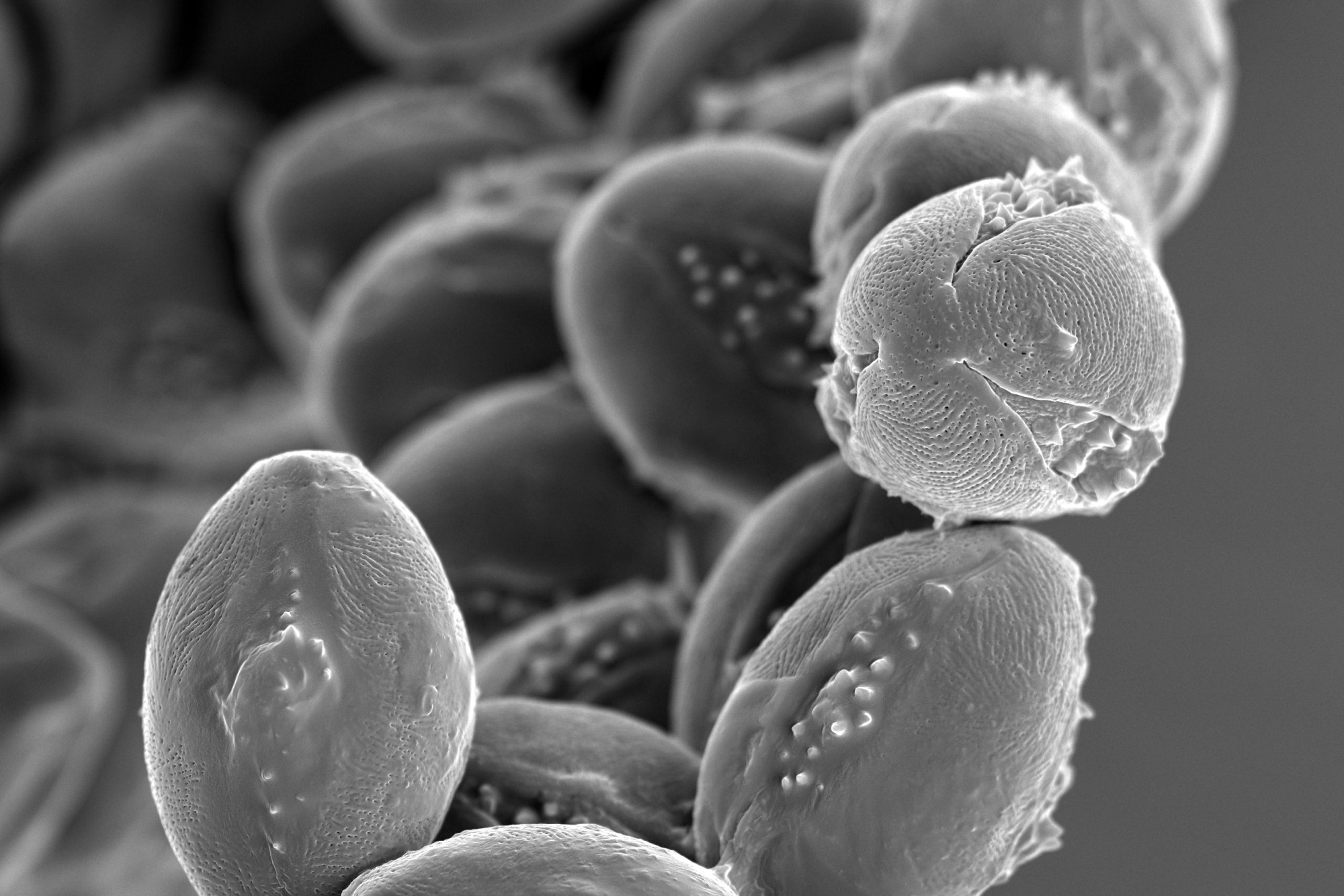
MACIEJ TRZASKOWSKI
Every chestnut has its thorn
The image shows chestnut tree pollen collected in May 2022 in Warsaw. The thorns protruding from the surface of pollen grains help the grains attach to insects that unknowingly carry them between trees.
The image was taken with the use of a Hitachi SU8230 scanning electron microscope owned by the Centre for Advanced Materials and Technologies CEZAMAT, Warsaw University of Technology, magnification: 2000X.


DIANA ALVES
A better world: seeding...
This is an SEM picture of zinc oxide synthesised under the scope of my research project. In the photograph we can see different structures with nanoflower shapes in different spaces and proportions, alluding to the blossoming of the process of becoming a better world. This better world - constantly changing - comprises the possibility of people and the planet thriving together.
The picture was taken at INL.


DIÓGENES PIVA
Union for a perfect sphere
Scanning electron microscopy (SEM) image of spherical structures obtained by the self-assembly of size-monodisperse polystyrene; INSTITUTION/INSTRUMENT: International Iberian Nanotechnology Laboratory (Braga)/ Quanta FEG 250 Field Emission SEM.


MACIEJ TRZASKOWSKI
Claws of the (micro)Beast
The image shows the tip of the leg of the larder beetle (Dermestes lardaceous). This beetle is a worldwide found pest in storage facilities. It can be found in cellars, wardrobes or magazines. It feeds mostly on animal-derived products such as lard or leather.
The image was taken with the use of a Hitachi SU8230 scanning electron microscope owned by the Centre for Advanced Materials and Technologies CEZAMAT, Warsaw University of Technology, magnification: 500X.


GEMMA RIUS
Screw it!
Tilted scanning electron microscope image of a self-standing Nanoscrew made out of silicon.
This Nanoscrew is a miniature (!) Probably the smallest on Earth: about 10 micrometres in length, while below 1 micrometre in diameter. It was obtained after applying a specialized, very anisotropic etching process, called reactive ion etching (RIE) at the Clean Room of the IMB-CNM-CSIC. The rippled shape of the threaded shank is also due to the processing conditions of the RIE, the so-called Bosch process.
The screw head (top particle) is the submicronic mask that was key to obtaining this high aspect ratio nanostructure.


RITA ALEXANDRE
The River Styx
Depicted is a SEM micrograph of resist patterned by nanoimprint lithography (NIL) on a silicon substrate, captured with NovaNanoSEM 650, at INL's clean room. In this area, the resist did not spread properly all over the substrate, resulting in the existence of vacant areas with a canal-like appearance.
This sample was patterned with a nanopillar structure with the goal of transferring this anti-reflective structure to other materials, such as the encapsulation glass of solar cells.

AGNES PURWIDYANTRI
Lend Me A Hand
The electron micrograph of polystyrene nanobeads holding hands with neighbouring particles after plasma treatment.
The photo was taken at Chang Gung University, Taiwan.

AGNES PURWIDYANTRI
Tropical Nanoparticles
The electron microscope image of transformation of CuO seed into CuO Nanoparticles during green-synthesis using L. citrus extracts precursor.
The photo was taken at the National Research and Innovation Agency of Indonesia (BRIN) Laboratory.
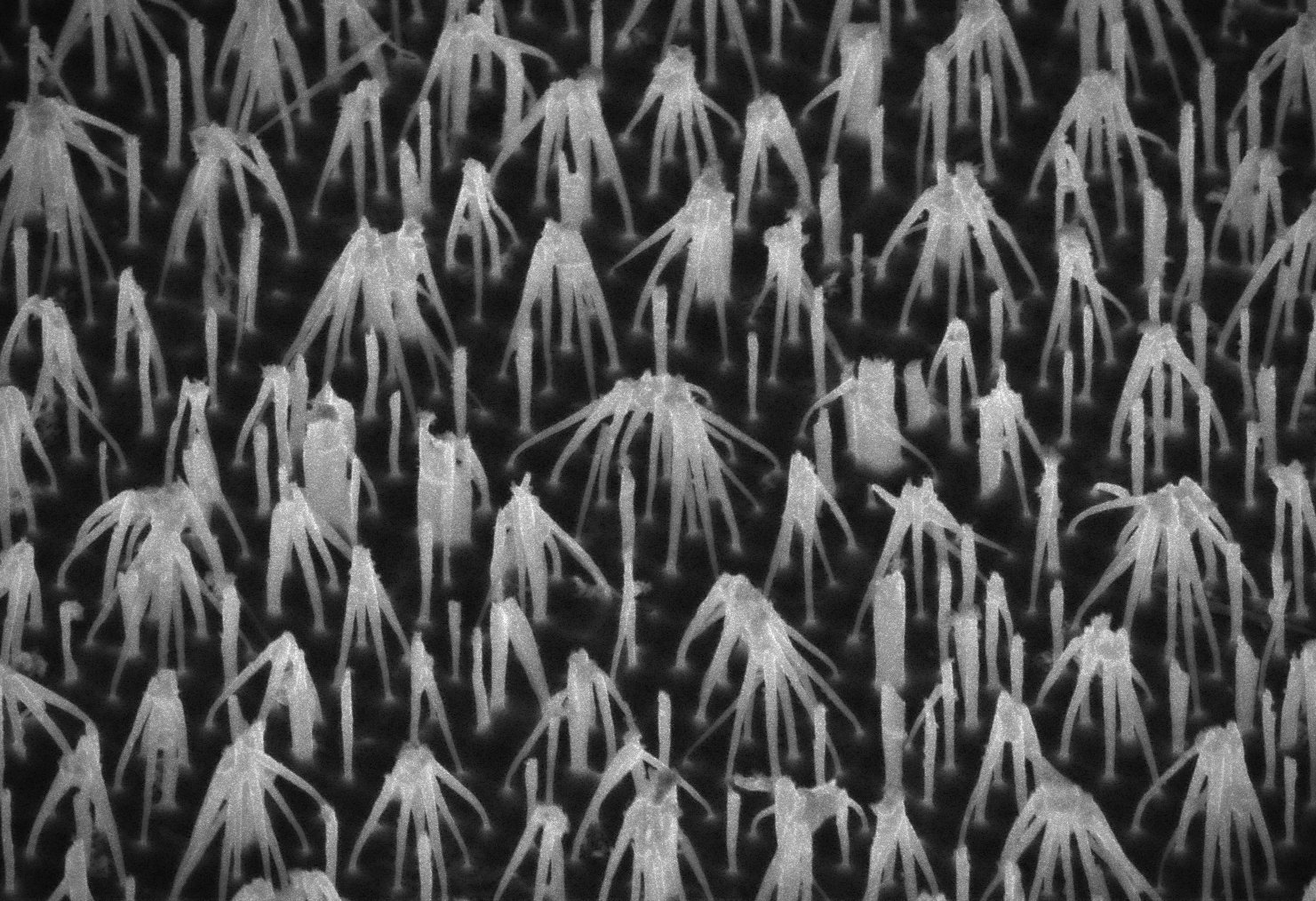
ALEXANDRE CHÍCHARO
Nano.Hugs.
This photo was taken with a scanning electron microscope (SEM) at INL. It portrays self-assembled gold and silicon nanopillars at the end of a nanofabrication process. Some of these nanopillars are companionless while others bend and hug each other. It was developed with Electron-beam lithography, a technique that uses a focused beam of electrons to draw custom shapes. The diameter of the nanopillars is between 50 and 150 nm and c.a. 1000 nm in height. While bulk materials are hard and brittle at these low sizes they are capable of bending without breaking.
This metamaterial is being used combined with 2D materials such as graphene to obtain surface plasmon polaritons that could be used as a new biosensor in the terahertz range. It is being developed in the framework of the GRAPHSENSE and TARGET projects.

ALEXANDRE CHÍCHARO
Nano.Active.Portals.
This photo was taken with a scanning electron microscope (SEM) at INL. It contains fine detail structures of gold nanoflowers at the end of a nanofabrication process. It was developed with Electron-beam lithography, a technique that uses a focused beam of electrons to draw custom shapes. The average diameter is 700nm and the inside hole diameters of c.a. 100nm.
Some of these 'nano portals' in this image are bright and some are dark as if some are active, particularly the centermost one. This charging effect comes from the electron cloud sharing between them.
This metamaterial is being used for the detection of tumor cells and metabolites in the framework of the BIOCELLPHE project. The technique being used are ultrasensitive Surface-enhanced Raman spectroscopy (SERS) and machine learning methods to unravel cancer biology.

ALEX DANTE
Dendrites of Hope I
The “Dendrites of Hope” photos were taken on July 14, 2022, at the INL cleanroom with a Nikon Elipse L200N optical microscope. It shows mysterious, marvellous tree branch-like dendrites of copper oxide grown on a copper layer over a silicon wafer.
This work is co-funded by the European Regional Development Fund (ERDF) through the Northern Regional Operational Program (NORTE 2020) of the Portugal 2020 Program [Project No. 45073, "ITEC Smart Automation I4.0"; Funding Reference: NORTE-01-0247-FEDER-045073.

ALEX DANTE
Dendrites of Hope II
The “Dendrites of Hope” photos were taken on July 14, 2022 at the INL cleanroom with a Nikon Elipse L200N optical microscope. It shows mysterious, marvellous tree branch-like dendrites of copper oxide grown on a copper layer over a silicon wafer.
This work is co-funded by the European Regional Development Fund (ERDF) through the Northern Regional Operational Program (NORTE 2020) of the Portugal 2020 Program [Project No. 45073, "ITEC Smart Automation I4.0"; Funding Reference: NORTE-01-0247-FEDER-045073.

ANA FRANCISCA CASTRO
Flea me to the Moon
The legs of a flea.
Photo taken in my Zoology classes with an optical microscope under the supervision of Dr Teresa Almeida.
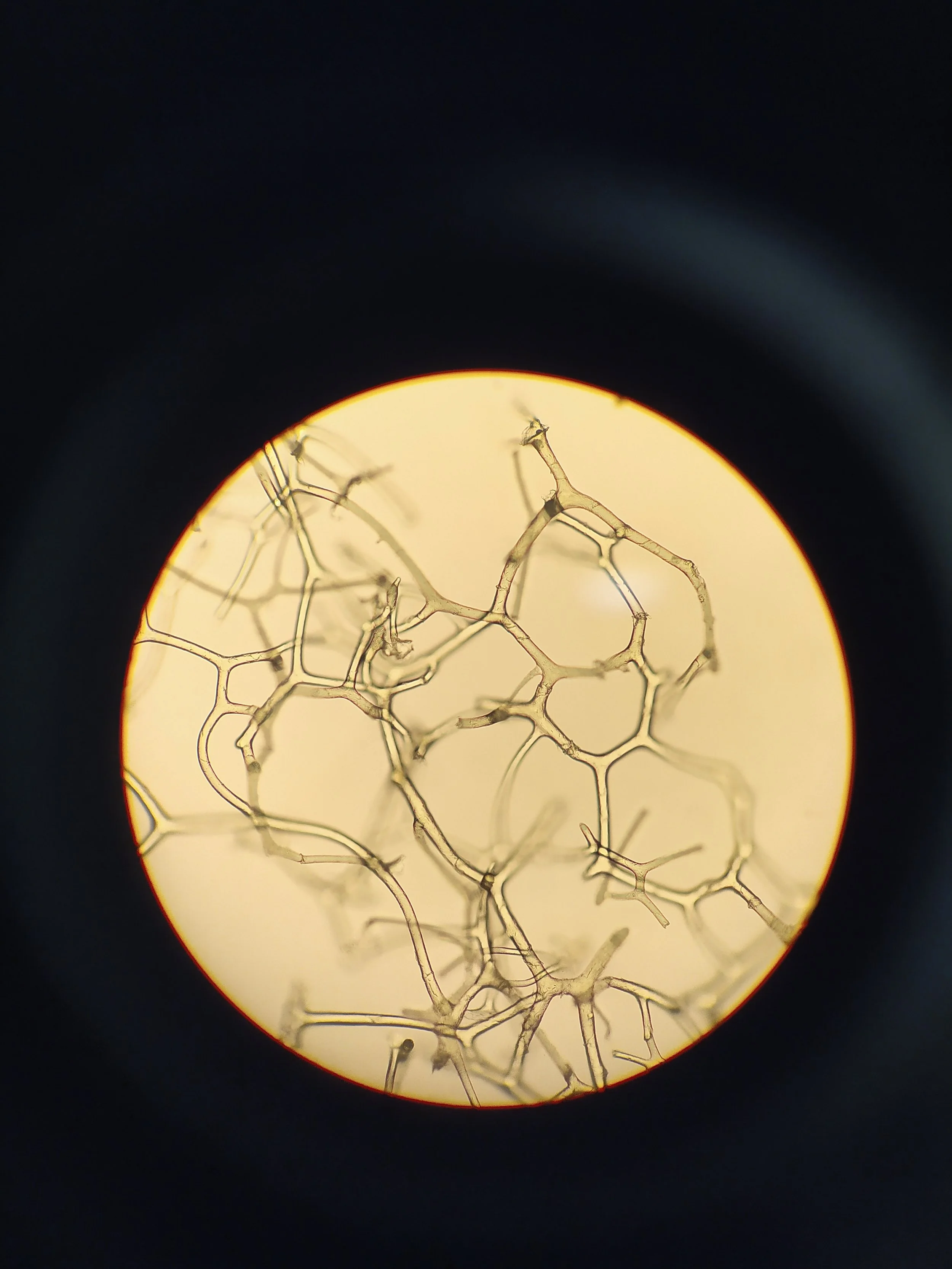
ANA FRANCISCA CASTRO
Sponge Skeleton
Spongin fibres (x100) of an individual of the most primitive group of animals: Porifera. Spongin is a protein which constitutes, together with the spicules, the sponge’s skeleton which has the function to protect and support the soft parts of these animals. Most of us are familiarized with sponges’ skeletons since they are used worldwide as bath sponges.
Photo taken in my Zoology classes with an optical microscope under the supervision of Dr Teresa Almeida.

PEDRO ANACLETO
There is always a rainbow somewhere, even behind a mistake.
The photo captures a sample defect (the tall dark thing that looks like a person) causing a non-uniform deposition of the silicon dioxide (SiO2) film on top of it. The various colours represent different SiO2 thicknesses, which naturally have a specific colour, thus creating a rainbow pattern.
I took this photo in the INL cleanroom using an optical microscope. The sample was used to fabricate micro solar cells.

PEDRO ANACLETO
Two worlds colliding
The photo captures a sample dipped into an electroplating solution to deposit Cu(In, Ga)Se2, a solar cell absorber material. The right-hand side of the sample was dipped into the electrodeposition solution (one world), while the left-hand side is where the metal contacts established an electrical connection (the other world).
I took this photo in the INL cleanroom using an optical microscope. The sample was used to fabricate micro solar cells.

ANDREA CAPASSO
Big worries for an incomparably small Sisyphus
Relativity did not just revolutionize the history of physics but widened the sight of humanity as a general concept. As individuals, we are centred on our inner selves, often thinking that our worries are bigger than others are, by their sheer size. In the photo, Sisyphus appears in his eternal struggle: having to roll an extremely heavy boulder up a hilltop, where he knows it will fall repeatedly, making his backbreaking effort an eternal one. This particular Sisyphus is shorter than a single mitochondrion, and the boulder would likely weigh as a couple of bacteria, but would you dare to tell him his struggle is whimsical? Once more, all is relative: only by keeping an open mind can we hope to prosper in harmony. (Graphene flakes with sub-micron lateral sizes were imaged with transmission electron microscopy at 100 keV accelerating voltage. The image was not corrected or modified after the acquisition).

ANDRÉ VIOLAS
NanoText Fonts 1
This photo was taken through the Scanning Electron Microscope (SEM). The T shape of the nanostructures is composed of two polymer resist films that were sequentially deposited. To obtain such nanostructures, we needed extensive process calibration and optimization to exploit the different properties of the two resist. The polymers were patterned using an industrial friend technique - NanoImprint Lithography, which is foreseen as a sustainable next-generation technology. Finally, these nanostructures will help in nanofabrication processes that will optimize the performance of ultrathin solar cells.

ANDRÉ VIOLAS
NanoText Fonts 2
This photo was taken through the Scanning Electron Microscope (SEM). The T shape of the nanostructures is composed of two polymers resist films that were sequentially deposited. To obtain such nanostructures, we needed extensive process calibration and optimization to exploit the different properties of the two resist. The polymers were patterned using an industrial friend technique - NanoImprint Lithography, which is foreseen as a sustainable next-generation technology. Finally, these nanostructures will help in nanofabrication processes that will optimize the performance of ultrathin solar cells.
With an increased SEM magnification, we can see how difficult may be to obtain such nanostructures, due to the required precision at very small sizes, i.e. at the nanoscale.
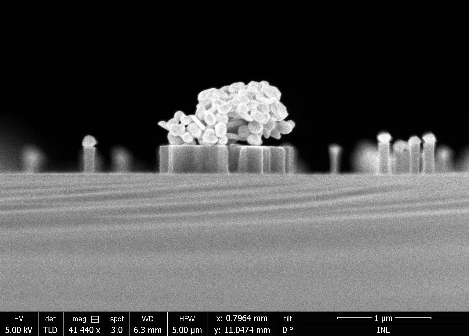
ANTÓNIO OLIVEIRA
Nanoparticles in an art gallery
This picture took in a scanning electron microscope that looks like stick-man are enjoying an art gallery, is actually nanoparticles with 150 nm (150*10-9 m) on top of Si pillars (or a table, in the case of the big cluster in the middle). The nanoparticles here actually act as a mask to etch the Si below.

ANTÓNIO OLIVEIRA
Nanoparticles try to recreate Europe
This picture was taken in a Scanning electron microscope has aggregates of very small nanoparticles (15 nm or 15*10-9 m) that resemble some Mediterranean countries. In the picture, you can see clearly the Iberian peninsula and the iconic Italy boot shape.

BEATRIZ CUSTÓDIO
Angry Wolf
Zebrafish telencephalon section with neurons (acetylated tubulin - yellow) and nuclei (Hoechst - cyan) stained, acquired under scanning confocal microscope.

BEATRIZ CUSTÓDIO
Bipolar zebrafish
Micro-CT acquisitions of zebrafish animals in different rotation positions.
The x-ray opacity of otoliths draws happy and angry faces depending on the rotation of the animal during micro-CT acquisition.

BRILIANT ADHI PRABOWO
Nanofurry Particles
SEM figure of semiconductor nanoparticles growth undergone acid treatments. The photo was taken at the National Research and Innovation Agency of Indonesia (BRIN).

BRILIANT ADHI PRABOWO
The Crater
The Nanosphere Lithography process generates a central micro-crater post-etching process.
The photo was taken at Chang Gung University, Taiwan.

BUSE NAZ GÜRBÜZ
Harmony of the needles
The image of oleogel constituted by sunflower oil, pea protein, glycerol monostearate and phytosterols under a microscope 20X (100μm).
This photo was taken in the INL facilities using NBI - Nikon Ni-E Upright Widefield/Fluorescence Microscope in the polarized mode.

BUSE NAZ GÜRBÜZ
Snowflake droplets
The image of oleogel constituted by sunflower oil, pea protein, glycerol monostearate and phytosterols under a microscope 20X (100μm).
This photo was taken in the INL facilities using NBI - Nikon Ni-E Upright Widefield/Fluorescence Microscope in the polarized mode.

CARINA CRUCHO
Beyond Silicon Valley
This is a transmission electron microscopy image (TEM) of pH-responsive silica-coated polystyrene nanoparticles. Silicon is, after oxygen, the most abundant element of the Earth’s crust and 75% of the Earth is made from silica – an oxide from silicon. The richness of silicon chemistry created almost unlimited opportunities for the development of environmentally benign nanomaterials and these smart coatings are the first of their kind.
The photo was taken at MicroLab/IST and I was surprised by the raspberry-like morphology.

DANIELA FIRMINO
MOFs - the porous crystalline frameworks from the future to solve our present problems
This photo is the perfect illustration of what a MOF sample usually looks like. It was taken in the lab, under UV lamp radiation. MOFs (Metal-Organic Frameworks) are highly organized porous coordination polymers and we can usually see a characteristic shine of these crystalline materials, even with the naked eye.

DANIEL BRITO
CIGS microtubes
CIGS is a semiconductor material used as an absorber layer for solar cells. The photo was taken in scanning electron microscopy (SEM).

DIANA ALVES
A little less narrowness, a little more blossoming, please!
This is a SEM picture of zinc oxide synthesised under the scope of my research project. I intended to produce this material with needle-like structures but instead, I end up with these nanoflowers. So, the title has a double meaning: this story behind it and also the need in this world to stop being small and narrow-minded and allow the opportunity to blossom. The photograph benefits from a wide negative space that contrasts with a space occupied by the flower- hence- the narrowing and the blossoming part.
The picture was taken at INL.

DIÓGENES PIVA
Green sphere destruction
Scanning electron microscopy (SEM) image of spherical structures obtained by the self-assembly of size-monodisperse polystyrene; INSTITUTION/INSTRUMENT: International Iberian Nanotechnology Laboratory (Braga)/ Quanta FEG 250 Field Emission SEM.

ELISABETE FERNANDES
The world needs love!
This photo is a Scanning Electron Microscope (SEM) image of bacteriophages immobilized on a gold (Au) surface. It was obtained by SEM at INL in 2012. I was searching for bacteriophages on the surface, and surprisingly I found a heart format.
At that time I named this photo “lovely phages”. Although, throughout time, this image more than ever resonates with me with an emergent need of our humanity: love!

FÁTIMA ZORRO
Above the clouds
"Above the clouds" is a scanning electron microscopy image of a ZnO surface taken at INL in a Dual Beam FIB-SEM.

FÁTIMA ZORRO
Haunted Pacman
"Haunted Pacman" is a transmission electron microscopy image of two ZnO microspheres. Those spheres were inserted in heating/biasing equipment and went through an experiment with high temperature and electric field, ending up partially deteriorated. The photo was taken at INL in a Double Corrected HRTEM - Titan Themis.

GEMMA RIUS
Nanopergamine
Scanning electron microscope image of an ultra-thin metal film.
This Nanopergamine is a only a few tens of nanometers thick (30nm), yet very robust. It was obtained upon a specialized delamination and transfer process of micropatterned metal films at the Clean Room of the IMB-CNM-CSIC.
The rolling is due to physical phenomena, including film stress and capillarity issues.

HELENA MACEDO
Wave of cells
This photo was taken with an optical microscope and depicts an H&E performed in an in vitro intestinal cellular model that I developed during my PhD at i3S, Porto (the place where the photo was taken).
In the photo, it is possible to see a layer of intestinal cells (dark blue) on top of a 3D collagen layer (light blue). The layer was supposed to be flat but something happened when cutting the sample and it got this shape. It reminded me of a sea wave and that's why I changed the colour from pink (the original H&E colour) to blue!
When I saw the topic of this year's ERN Photo Competition I immediately thought of this photo for 2 reasons: it shows an in vitro intestinal model that contributes to reducing the number of animal experiments in scientific research and it reminds us of the sea, an extremely valuable asset that we need to protect!

IHSAN ÇAHA
Microstructure of Ti-12Nb alloy processed by powder metallurgy
The raw materials used to produce Ti-12Nb alloy were TiH2 and Nb powders. The alloy produced by powder metallurgy, TiH2 and Nb powders were homogenized by mixing for 1 h in a Turbula® multidirectional mixer. Green compacts were acquired under 700 MPa pressure using zinc stearate as a die wall lubricant. The samples were sintered in a tubular furnace under a high vacuum (10−5 mbar) by holding 4 h at 1250 °C.
The microstructure of the alloys was characterized by optical microscopy (Leica, DM2500) after preparing the samples by grinding down to 1200 grit SiC paper, then polished with colloidal silica suspension (0.02 μm particle size), and etched by Kroll’s reagent (3 mL HF, 6 mL HNO3, and 91 mL H2O). High magnification.

IHSAN ÇAHA
Microstructure of Ti-12Nb alloy processed by powder metallurgy
The raw materials used to produce Ti-12Nb alloy were TiH2 and Nb powders. The alloy produced by powder metallurgy, TiH2 and Nb powders were homogenized by mixing for 1 h in a Turbula® multidirectional mixer. Green compacts were acquired under 700 MPa pressure using zinc stearate as a die-wall lubricant. The samples were sintered in a tubular furnace under a high vacuum (10−5 mbar) by holding 4 h at 1250 °C.
The microstructure of the alloys was characterized by optical microscopy (Leica, DM2500) after preparing the samples by grinding down to 1200 grit SiC paper, then polished with colloidal silica suspension (0.02 μm particle size), and etched by Kroll’s reagent (3 mL HF, 6 mL HNO3, and 91 mL H2O). Low magnification.

JOÃO CUNHA
Microscale Tetris: Detail of a Holographic Mask
This Scanning Electron Microscope image shows a detail of a holographic mask consisting of a gold-coated photoresist whose topographic features have been defined through grayscale lithography. Holographic masks allow to encode visual images which are revealed only when shining light on the mask. The topographic features contain the information of the hologram's image in an apparently random succession of rectangular shapes with several heights (multilevel) that appear to have no connection with each other or with the encoded image.
In an abstract way, the mask's multilevel features seem like pieces of a microscale Tetris game (white bar corresponding to 10 micrometres, around 10 times smaller than a human hair).
This micrograph was taken at the International Iberian Nanotechnology (INL) Micro and Nano Fabrication facility using a Scanning Electron Microscope FEI NovaNanoSEM 650.

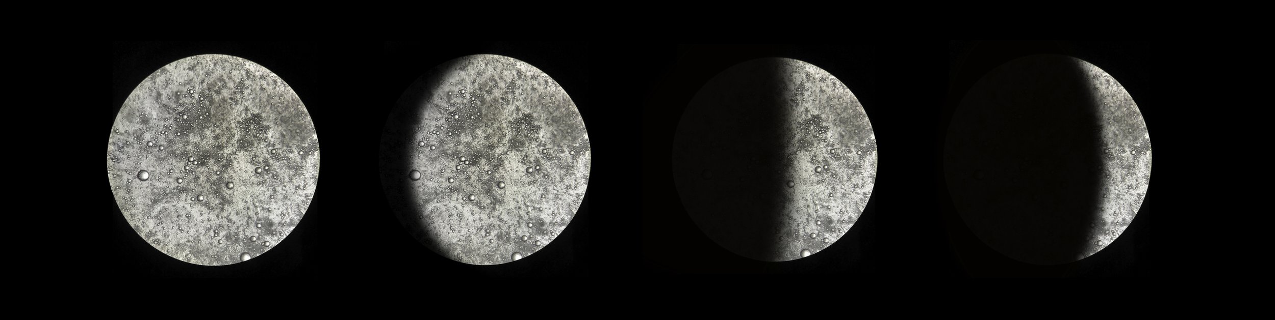
JOÃO MARTINHO MOURA
Sci-fi Miners. Performance at Le Centquatre, Paris, France, 2020
Generated reconstruction calculated by SEM microscopy observations at INL cleanroom, representing nanoparticle foam to produce clean hydrogen fuel.
Approximate scale: 10µm
Picture from recordings of João Martinho Moura’s virtual reality performance at Le Centquatre, on 28 February 2020, in Paris. STARTS event from the European Commission. Imagine one millimetre… now, divide it by one million. That is one nanometer, an incredibly small-scale unit where Sci-fi Miners’ narrative happens, a journey beginning at the millimetre scale (1mm), ending at the size of one nanometer (1nm), employing virtual reality technologies.
An audio-visual, interactive, and virtual reality performance where the artist takes the audience on a journey on the nanometric scale of matter. Work designed by João Martinho Moura with the support of the STARTS Residencies as part of the STARTS program of the European Commission, based on the technological elements of the CritCat Project (INL – International Iberian Nanotechnology Laboratory and Aalto University).

JOSÉ GAMA
Light diffraction effect of Silica Nanowrinkles on PDMS
This phone camera image was taken with Xiaomi Redmi Note 8T. The light diffraction effect is noticeable as a clear colour gradient on the PDMS sample surface.

JULIANA SOUSA
Two different materials but not too different
The image was obtained by scanning electron microscope, where we can see two different types of fibres (carbon and cerium fibres). These materials were prepared using the electrospinning technique and were used in the degradation of inorganic pollutants present in rivers. Using two different precursors obtained materials with different chemical proprieties. The synergy between the two makes them an excellent catalyst, where the weakness of one is compensated by the other. The catalyst presents excellent catalytic properties and high stability in the degradation of the pollutants present in water.

MAFALDA NETO
Shape-shifter
This primary human intestinal organoid skipped a beat and is now morphing into an untamed heart.
(Image acquired in Zeiss LSM 780 Confocal Microscope; Red: E-Cadherin, Blue: Nuclei)

MANUEL RODRÍGUEZ OSORIO
E. coli after a night on a bed of nails
This photo was taken on a sample of nanoimprinted hard PDMS material. The pattern replicates the moth-eye nanocones, which are known to have antibacterial properties. Bacteria try to adhere to the nanocones, and so the wall is deformed accordingly. These two bacteria were flipped by using scotch tape in order to show the indentations. The picture was taken using a Carl Zeiss Auriga CrossBeam Workstation (Instituto IMDEA Nanociencia, Madrid).

MARÍA TERESA ALAMEDA FELGUEIRAS
Bringing the damage to light
Stereometric reconstruction of a flipped-up E. coli bacteria based on Scanning Electron Microscopy images taken in a Carl Zeiss AURIGA CrossBeam Workstation for measuring damage inflicted by Moth-eye nanocones fabricated on PDMS substrate. More information about this image processing can be found here.

MARÍA TERESA ALAMEDA FELGUEIRAS
Cellular love breakup
Image of MCF7 cells growing on a hierarchical topography comprising micro-gratings and nanocones fabricated on PDMS substrate taken in a Scanning Electron Microscopy Carl Zeiss EVO HD15.

MIGUEL XAVIER
Skin cancer cells under division
This photo captured skin cancer cells undergoing mitosis when grown in lab-based cell culture conditions. They are used in organ-on-a-chip platforms to establish sustainable models for in vitro research that reduce the need for animal-based studies.
The photo was taken using a Zeiss LSM780 confocal laser scanning microscope at the International Iberian Nanotechnology Laboratory.

PATRÍCIA RODRIGUES
Staggered Herringbone Mixer
The fluid dynamics is closely related to the scaling effect. At the macroscale, the fluids follow the turbulent flow, a term also associated with chaotic flow, where the fluids move in all directions. For a better understanding, let's suppose our blood flow in the arteries. A curve in the aorta leads to a bend in the blood flow, which causes the blood cells to get mixed with each other. Sometimes a bulge in the arteries also induces a turbulent blood flow.
Interestingly, when dealing with small channels (dimensions that can go from tens to hundreds of micrometres), the fluids exhibit a distinct behaviour from what happens in "real life". At the micro-scale, two different fluids flowing side-by-side do not mix and, therefore, move parallel to each other along the micro-channel. This control of fluids can bring advantages in some cases; however, it can become a problem when the mixing of fluids is of utmost importance.
In microfluidic, disruption of the fluid flow can be achieved by incorporating repeated patterns of grooves on the bottom of the micro-channel. These structures will induce helical motions of the moving fluids, thereby improving the mixing efficiency.
Here, the confocal image shows the performance of the staggered herringbone mixer (SHM) by mixing 5 uM of fluorescein and water (0 uM). Right away after introducing them in the micro-channel, the two fluids stretch and fold into another leading to a complete mixing before the end of the micro-channel. This ensured that a microfluidic system can be used for experimental studies where mixing of different solutions are required in small volumes.

PATRICIA TALADRIZ-BLANCO
Searching for nanoplastics in the sea
Scanning electron microscopy micrograph of water from the North Sea taken with an FEI Quanta 650 microscope. The micrograph shows the different salts present in the seawater and possible nanoplastic particles attached to them.

RITA ALEXANDRE
The Dark Forest
Silicon nanopillars fabricated by Reactive Ion Etch with gold nanoparticles as the etch mask, captured with NovaNanoSEM 650, at INL's clean room. This nanostructure exhibits broadband anti-reflection, which can be employed to boost the performance of solar cells once imprinted in the encapsulation glass, for instance.

RODICA VOICU
Cracks but not cracks
Cracks in the gold nano-layer deposited on SU-8 polymer during the processing of a microgripper device. Photo at the microscope in the technological department, IMT Bucharest.

RODICA VOICU
Diamond in the lab
Under the microscope, tonner microparticle of carbon on SU-8 surface. In IMT Bucharest technological laboratory.

SANDRA AMARAL
One small step for a particle, a giant leap for sustainability.
Fragrance microcapsules are prepared from biomaterials, designed to be sustainable and biodegradable, and potentially substitute their plastic counterparts. Image is taken from the eyepiece of a microscope. Institution/instrument: International Iberian Nanotechnology Laboratory (INL)/ Nikon SMZ1500 Stereoscopic Zoom microscope.
SANDRA AMARAL
Fading away
Fragrance microcapsules are prepared from biomaterials, designed to be sustainable and biodegradable, and potentially substitute their plastic
counterparts. Images were taken from the eyepiece of a microscope from different angles and composed with Photoshop. The photo represents the inability of overcoming the sustainability issues to fight climate change. Institution/instrument: International Iberian Nanotechnology Laboratory (INL)/ Nikon SMZ1500 Stereoscopic Zoom microscope.